{"title":"Fixation methods can differentially affect ciliary protein immunolabeling.","authors":"Kiet Hua, Russell J Ferland","doi":"10.1186/s13630-017-0045-9","DOIUrl":null,"url":null,"abstract":"<p><strong>Background: </strong>Primary cilia are immotile, microtubule-based organelles present on most cells. Defects in primary cilia presence/function result in a category of developmental diseases referred to as ciliopathies. As the cilia field progresses, there is a need to consider both the ciliary and extraciliary roles of cilia proteins. However, traditional fixation methods are not always suitable for examining the full range of localizations of cilia proteins. Here, we tested a variety of fixation methods with commonly used cilia markers to determine the most appropriate fixation method for different cilia proteins.</p><p><strong>Methods: </strong>Mouse inner medullary collecting duct and human retinal pigmented epithelial cells were grown to confluence, serum starved, and fixed with one of the following fixation agents: paraformaldehyde-sucrose, paraformaldehyde-PBS, methanol, cytoskeletal buffer followed by methanol, or three variations of cytoskeletal buffer-paraformaldehyde fixation. Each cell type and fixation method combination was probed with the following ciliary markers: acetylated α-tubulin, detyrosinated tubulin, polyglutamylated tubulin, β-tubulin, adenylyl cyclase 3 (AC3), ADP-ribosylation factor-like protein 13b (Arl13b), centrosome and spindle pole associated protein 1 (CSPP1), or intraflagellar transport protein 20 (IFT20). Intraflagellar transport protein 88 (IFT88) and GM130 (Golgi marker) were also used. We assessed actin (via phalloidin) and microtubule integrity, centrioles, cilia, and two extraciliary sites (mitotic figures and Golgi).</p><p><strong>Results: </strong>For the cilia markers examined, paraformaldehyde fixation preserved cilia immunolabeling of cilia-membrane proteins (AC3 and Arl13b), but failed to reveal cilia immunostaining of axonemal proteins (CSPP1 and IFT20). Methanol revealed cilia labeling for some axonemal proteins, but not others, and this depended on cell type. Generally, any method that first included a wash in cytoskeletal buffer, before fixing, revealed more distinct cilia immunolabeling for axonemal proteins (CSPP1, IFT20, and IFT88), but resulted in the loss of cilia labeling for cilia-membrane proteins (AC3 and Arl13b). All three different post-translational modifications of tubulin antibodies positively immunolabeled cilia in all fixation methods tested. Ultimately, we found that fixing cells in a solution of paraformaldehyde prepared in cytoskeletal buffer allowed for the preservation of cilia immunolabeling for most cilia proteins tested and allowed visualization of two extraciliary sites (mitotic figures and Golgi).</p><p><strong>Conclusion: </strong>Some general patterns were observed to guide in the choice of a fixation agent. Cilia-membrane proteins generally benefit from quick fixation with no prior permeabilization, whereas axonemal proteins tend to benefit from permeabilization and use of cytoskeletal buffer.</p>","PeriodicalId":38134,"journal":{"name":"Cilia","volume":" ","pages":"5"},"PeriodicalIF":0.0000,"publicationDate":"2017-03-24","publicationTypes":"Journal Article","fieldsOfStudy":null,"isOpenAccess":false,"openAccessPdf":"https://sci-hub-pdf.com/10.1186/s13630-017-0045-9","citationCount":"45","resultStr":null,"platform":"Semanticscholar","paperid":null,"PeriodicalName":"Cilia","FirstCategoryId":"1085","ListUrlMain":"https://doi.org/10.1186/s13630-017-0045-9","RegionNum":0,"RegionCategory":null,"ArticlePicture":[],"TitleCN":null,"AbstractTextCN":null,"PMCID":null,"EPubDate":"2017/1/1 0:00:00","PubModel":"eCollection","JCR":"Q2","JCRName":"Biochemistry, Genetics and Molecular Biology","Score":null,"Total":0}
引用次数: 45
Abstract
Background: Primary cilia are immotile, microtubule-based organelles present on most cells. Defects in primary cilia presence/function result in a category of developmental diseases referred to as ciliopathies. As the cilia field progresses, there is a need to consider both the ciliary and extraciliary roles of cilia proteins. However, traditional fixation methods are not always suitable for examining the full range of localizations of cilia proteins. Here, we tested a variety of fixation methods with commonly used cilia markers to determine the most appropriate fixation method for different cilia proteins.
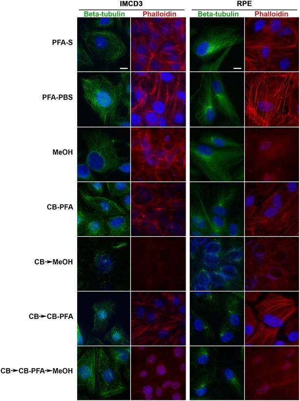
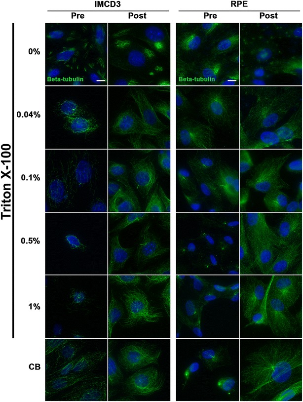
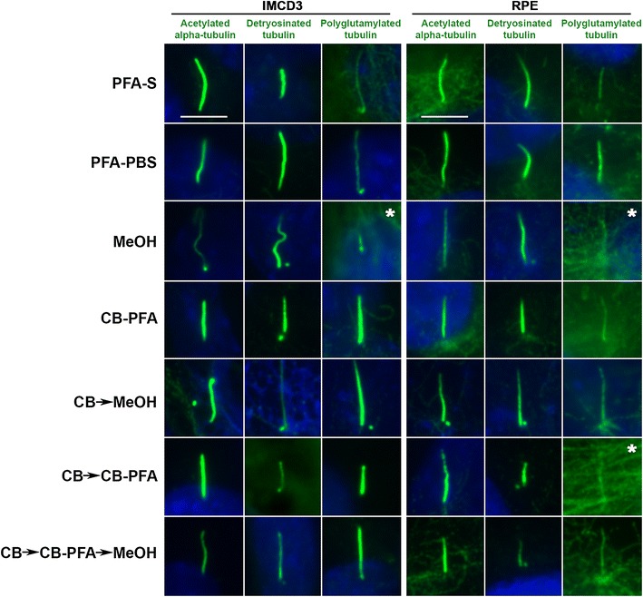
Methods: Mouse inner medullary collecting duct and human retinal pigmented epithelial cells were grown to confluence, serum starved, and fixed with one of the following fixation agents: paraformaldehyde-sucrose, paraformaldehyde-PBS, methanol, cytoskeletal buffer followed by methanol, or three variations of cytoskeletal buffer-paraformaldehyde fixation. Each cell type and fixation method combination was probed with the following ciliary markers: acetylated α-tubulin, detyrosinated tubulin, polyglutamylated tubulin, β-tubulin, adenylyl cyclase 3 (AC3), ADP-ribosylation factor-like protein 13b (Arl13b), centrosome and spindle pole associated protein 1 (CSPP1), or intraflagellar transport protein 20 (IFT20). Intraflagellar transport protein 88 (IFT88) and GM130 (Golgi marker) were also used. We assessed actin (via phalloidin) and microtubule integrity, centrioles, cilia, and two extraciliary sites (mitotic figures and Golgi).
Results: For the cilia markers examined, paraformaldehyde fixation preserved cilia immunolabeling of cilia-membrane proteins (AC3 and Arl13b), but failed to reveal cilia immunostaining of axonemal proteins (CSPP1 and IFT20). Methanol revealed cilia labeling for some axonemal proteins, but not others, and this depended on cell type. Generally, any method that first included a wash in cytoskeletal buffer, before fixing, revealed more distinct cilia immunolabeling for axonemal proteins (CSPP1, IFT20, and IFT88), but resulted in the loss of cilia labeling for cilia-membrane proteins (AC3 and Arl13b). All three different post-translational modifications of tubulin antibodies positively immunolabeled cilia in all fixation methods tested. Ultimately, we found that fixing cells in a solution of paraformaldehyde prepared in cytoskeletal buffer allowed for the preservation of cilia immunolabeling for most cilia proteins tested and allowed visualization of two extraciliary sites (mitotic figures and Golgi).
Conclusion: Some general patterns were observed to guide in the choice of a fixation agent. Cilia-membrane proteins generally benefit from quick fixation with no prior permeabilization, whereas axonemal proteins tend to benefit from permeabilization and use of cytoskeletal buffer.


 分享
分享
 求助内容:
求助内容: 应助结果提醒方式:
 扫码关注我们
