{"title":"Bone marker gene expression in calvarial bones: different bone microenvironments.","authors":"Osama Al-Amer","doi":"10.1186/s40709-017-0066-y","DOIUrl":null,"url":null,"abstract":"<p><strong>Background: </strong>In calvarial mice, mesenchymal stem cells (MSCs) differentiate into osteoprogenitor cells and then differentiate into osteoblasts that differentiate into osteocytes, which become embedded within the bone matrix. In this case, the cells participating in bone formation include MSCs, osteoprogenitor cells, osteoblasts and osteocytes. The calvariae of C57BL/KaLwRijHsD mice consist of the following five bones: two frontal bones, two parietal bones and one interparietal bone. This study aimed to analyse some bone marker genes and bone related genes to determine whether these calvarial bones have different bone microenvironments.</p><p><strong>Methods: </strong>C57BL/KaLwRijHsD calvariae were carefully excised from five male mice that were 4-6 weeks of age. Frontal, parietal, and interparietal bones were dissected to determine the bone microenvironment in calvariae. Haematoxylin and eosin staining was used to determine the morphology of different calvarial bones under microscopy. TaqMan was used to analyse the relative expression of Runx2, OC, OSX, RANK, RANKL, OPG, N-cadherin, E-cadherin, FGF2 and FGFR1 genes in different parts of the calvariae.</p><p><strong>Results: </strong>Histological analysis demonstrated different bone marrow (BM) areas between the different parts of the calvariae. The data show that parietal bones have the smallest BM area compared to frontal and interparietal bones. TaqMan data show a significant increase in the expression level of Runx2, OC, OSX, RANKL, OPG, FGF2 and FGFR1 genes in the parietal bones compared with the frontal and interparietal bones of calvariae.</p><p><strong>Conclusion: </strong>This study provides evidence that different calvarial bones, frontal, parietal and interparietal, contain different bone microenvironments.</p>","PeriodicalId":87292,"journal":{"name":"","volume":"24 ","pages":"9"},"PeriodicalIF":0.0,"publicationDate":"2017-05-16","publicationTypes":"Journal Article","fieldsOfStudy":null,"isOpenAccess":false,"openAccessPdf":"https://sci-hub-pdf.com/10.1186/s40709-017-0066-y","citationCount":"5","resultStr":null,"platform":"Semanticscholar","paperid":null,"PeriodicalName":"","FirstCategoryId":"99","ListUrlMain":"https://doi.org/10.1186/s40709-017-0066-y","RegionNum":0,"RegionCategory":null,"ArticlePicture":[],"TitleCN":null,"AbstractTextCN":null,"PMCID":null,"EPubDate":"2017/12/1 0:00:00","PubModel":"eCollection","JCR":"","JCRName":"","Score":null,"Total":0}
引用次数: 5
Abstract
Background: In calvarial mice, mesenchymal stem cells (MSCs) differentiate into osteoprogenitor cells and then differentiate into osteoblasts that differentiate into osteocytes, which become embedded within the bone matrix. In this case, the cells participating in bone formation include MSCs, osteoprogenitor cells, osteoblasts and osteocytes. The calvariae of C57BL/KaLwRijHsD mice consist of the following five bones: two frontal bones, two parietal bones and one interparietal bone. This study aimed to analyse some bone marker genes and bone related genes to determine whether these calvarial bones have different bone microenvironments.
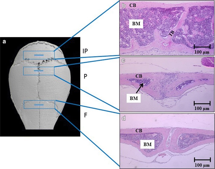
Methods: C57BL/KaLwRijHsD calvariae were carefully excised from five male mice that were 4-6 weeks of age. Frontal, parietal, and interparietal bones were dissected to determine the bone microenvironment in calvariae. Haematoxylin and eosin staining was used to determine the morphology of different calvarial bones under microscopy. TaqMan was used to analyse the relative expression of Runx2, OC, OSX, RANK, RANKL, OPG, N-cadherin, E-cadherin, FGF2 and FGFR1 genes in different parts of the calvariae.
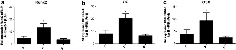
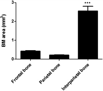
Results: Histological analysis demonstrated different bone marrow (BM) areas between the different parts of the calvariae. The data show that parietal bones have the smallest BM area compared to frontal and interparietal bones. TaqMan data show a significant increase in the expression level of Runx2, OC, OSX, RANKL, OPG, FGF2 and FGFR1 genes in the parietal bones compared with the frontal and interparietal bones of calvariae.
Conclusion: This study provides evidence that different calvarial bones, frontal, parietal and interparietal, contain different bone microenvironments.


 分享
分享
 求助内容:
求助内容: 应助结果提醒方式:
 扫码关注我们
