Anni Wong, Richard Chan Woo Park, Neena M Mirani, Jean Anderson Eloy
{"title":"Myxofibrosarcoma of the maxillary sinus.","authors":"Anni Wong, Richard Chan Woo Park, Neena M Mirani, Jean Anderson Eloy","doi":"10.2500/ar.2017.8.0200","DOIUrl":null,"url":null,"abstract":"<p><strong>Background: </strong>Myxofibrosarcoma (MFS) is a common sarcoma in the extremities of older individuals but is extremely uncommon in the head and neck region. Diagnosis may be challenging but is critical to the management of the patient. We discuss the radiographic and histopathologic characteristics of this destructive tumor. The distinguishing features of MFS and its differential diagnosis are reviewed to familiarize the managing otolaryngologist with this rare entity.</p><p><strong>Methods: </strong>A 61-year-old woman presents with a 6-week history of severe left facial pain and left eye pain. Imaging demonstrates significant right and left-sided maxillary sinus opacification with destruction of the left maxillary sinus as well as the left medial and inferior orbital walls.</p><p><strong>Results: </strong>Histopathologic examination revealed spindle and stellate tumor cells of variable cellularity in myxoid stroma with cellular pleomorphism consistent with MFS of intermediate-to high grade. The patient underwent resection of the left-sided lesion with orbital exenteration and repair of the defect with microvascular free flap followed by postoperative radiotherapy.</p><p><strong>Conclusion: </strong>MFS must be differentiated from other lesions with myxoid qualities. Histopathologic examination is required for definitive diagnosis. Management includes complete tumor excision with adequate tumor margins. Adjuvant postoperative radiotherapy should be considered for larger tumors with positive resection margins or lesions of intermediate-to-high grade.</p>","PeriodicalId":45192,"journal":{"name":"Allergy & Rhinology","volume":"8 2","pages":"95-99"},"PeriodicalIF":1.2000,"publicationDate":"2017-06-01","publicationTypes":"Journal Article","fieldsOfStudy":null,"isOpenAccess":false,"openAccessPdf":"https://sci-hub-pdf.com/10.2500/ar.2017.8.0200","citationCount":"7","resultStr":null,"platform":"Semanticscholar","paperid":null,"PeriodicalName":"Allergy & Rhinology","FirstCategoryId":"1085","ListUrlMain":"https://doi.org/10.2500/ar.2017.8.0200","RegionNum":0,"RegionCategory":null,"ArticlePicture":[],"TitleCN":null,"AbstractTextCN":null,"PMCID":null,"EPubDate":"","PubModel":"","JCR":"Q1","JCRName":"OTORHINOLARYNGOLOGY","Score":null,"Total":0}
引用次数: 7
Abstract
Background: Myxofibrosarcoma (MFS) is a common sarcoma in the extremities of older individuals but is extremely uncommon in the head and neck region. Diagnosis may be challenging but is critical to the management of the patient. We discuss the radiographic and histopathologic characteristics of this destructive tumor. The distinguishing features of MFS and its differential diagnosis are reviewed to familiarize the managing otolaryngologist with this rare entity.
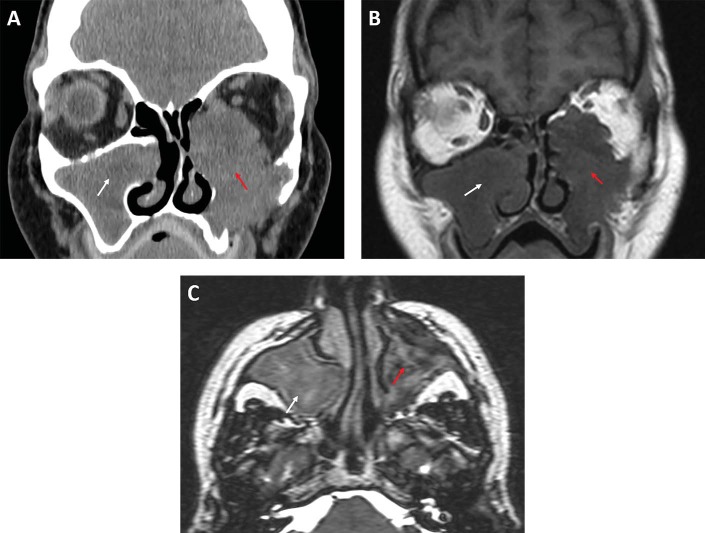
Methods: A 61-year-old woman presents with a 6-week history of severe left facial pain and left eye pain. Imaging demonstrates significant right and left-sided maxillary sinus opacification with destruction of the left maxillary sinus as well as the left medial and inferior orbital walls.
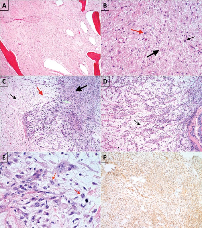
Results: Histopathologic examination revealed spindle and stellate tumor cells of variable cellularity in myxoid stroma with cellular pleomorphism consistent with MFS of intermediate-to high grade. The patient underwent resection of the left-sided lesion with orbital exenteration and repair of the defect with microvascular free flap followed by postoperative radiotherapy.
Conclusion: MFS must be differentiated from other lesions with myxoid qualities. Histopathologic examination is required for definitive diagnosis. Management includes complete tumor excision with adequate tumor margins. Adjuvant postoperative radiotherapy should be considered for larger tumors with positive resection margins or lesions of intermediate-to-high grade.


 分享
分享
 求助内容:
求助内容: 应助结果提醒方式:
 扫码关注我们
