Luciano A Favorito, Suelen F Costa, Waldemar S Costa, Rodrigo Vieiralves, Fabio O Bernardo, Francisco J B Sampaio
{"title":"Study of Testicular Structure in Fetuses with Prune Belly Syndrome.","authors":"Luciano A Favorito, Suelen F Costa, Waldemar S Costa, Rodrigo Vieiralves, Fabio O Bernardo, Francisco J B Sampaio","doi":"10.1155/2017/3254980","DOIUrl":null,"url":null,"abstract":"<p><strong>Purpose: </strong>To compare the structure of the testis in fetuses with prune belly syndrome (PBS) to normal controls.</p><p><strong>Materials and methods: </strong>We studied 6 testes obtained from 3 fetuses with PBS and 14 testes from 7 male fetuses. The testicular specimens were cut into 5-<i>μ</i>m thick sections and stained with hematoxylin and eosin (HE), to observe the seminiferous tubules; Weigert's solution to observe elastic fibers; and picrosirius red to observe collagen. The images were captured with an Olympus BX51 microscope and Olympus DP70 camera. The stereological analysis was done with the Image Pro and Image J programs. Means were statistically compared using the Mann-Whitney <i>U</i> test (<i>p</i> < 0.005).</p><p><strong>Results: </strong>Quantitative analysis documented no differences (<i>p</i> = 0.4) in number of seminiferous tubules (ST) in PBS testes (mean = 8.87%, SD = 1.59), when compared to the control (mean = 11.4%, SD = 2.99) and no differences (<i>p</i> = 0.8) in diameter of ST in PBS testes (mean = 52.85 <i>μ</i>m, SD = 1.58) when compared to the control group (mean = 53.17 <i>μ</i>m, SD = 1.55), but we did observe a lower number (<i>p</i> = 0.0002) of Leydig cells in the PBS testes (mean = 67.03% and SD = 3.697) when compared to the control group (mean = 90.1% and SD = 2.986).</p><p><strong>Conclusions: </strong>Our study showed a lower concentration of Leydig cells in the triad syndrome fetuses.</p>","PeriodicalId":7490,"journal":{"name":"Advances in Urology","volume":"2017 ","pages":"3254980"},"PeriodicalIF":2.3000,"publicationDate":"2017-01-01","publicationTypes":"Journal Article","fieldsOfStudy":null,"isOpenAccess":false,"openAccessPdf":"https://sci-hub-pdf.com/10.1155/2017/3254980","citationCount":"7","resultStr":null,"platform":"Semanticscholar","paperid":null,"PeriodicalName":"Advances in Urology","FirstCategoryId":"1085","ListUrlMain":"https://doi.org/10.1155/2017/3254980","RegionNum":0,"RegionCategory":null,"ArticlePicture":[],"TitleCN":null,"AbstractTextCN":null,"PMCID":null,"EPubDate":"2017/5/21 0:00:00","PubModel":"Epub","JCR":"Q3","JCRName":"UROLOGY & NEPHROLOGY","Score":null,"Total":0}
引用次数: 7
Abstract
Purpose: To compare the structure of the testis in fetuses with prune belly syndrome (PBS) to normal controls.
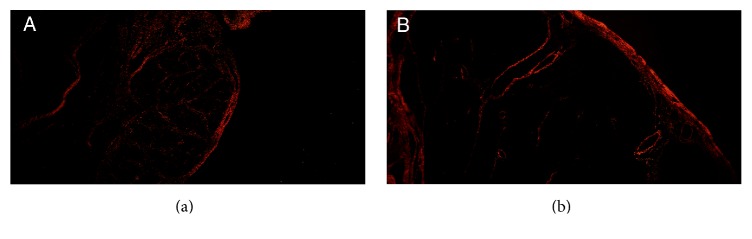
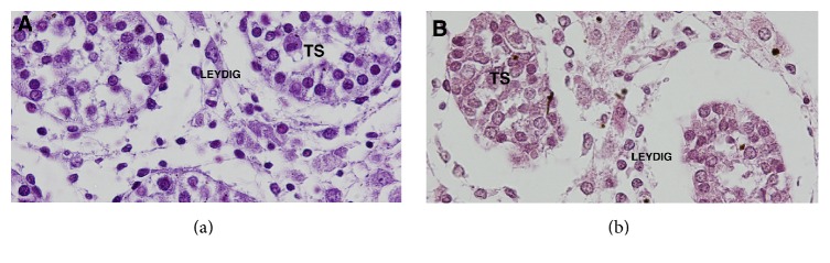
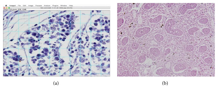
Materials and methods: We studied 6 testes obtained from 3 fetuses with PBS and 14 testes from 7 male fetuses. The testicular specimens were cut into 5-μm thick sections and stained with hematoxylin and eosin (HE), to observe the seminiferous tubules; Weigert's solution to observe elastic fibers; and picrosirius red to observe collagen. The images were captured with an Olympus BX51 microscope and Olympus DP70 camera. The stereological analysis was done with the Image Pro and Image J programs. Means were statistically compared using the Mann-Whitney U test (p < 0.005).
Results: Quantitative analysis documented no differences (p = 0.4) in number of seminiferous tubules (ST) in PBS testes (mean = 8.87%, SD = 1.59), when compared to the control (mean = 11.4%, SD = 2.99) and no differences (p = 0.8) in diameter of ST in PBS testes (mean = 52.85 μm, SD = 1.58) when compared to the control group (mean = 53.17 μm, SD = 1.55), but we did observe a lower number (p = 0.0002) of Leydig cells in the PBS testes (mean = 67.03% and SD = 3.697) when compared to the control group (mean = 90.1% and SD = 2.986).
Conclusions: Our study showed a lower concentration of Leydig cells in the triad syndrome fetuses.
期刊介绍:
Advances in Urology is a peer-reviewed, open access journal that publishes state-of-the-art reviews and original research papers of wide interest in all fields of urology. The journal strives to provide publication of important manuscripts to the widest possible audience worldwide, without the constraints of expensive, hard-to-access, traditional bound journals. Advances in Urology is designed to improve publication access of both well-established urologic scientists and less well-established writers, by allowing interested scientists worldwide to participate fully.


 分享
分享
 求助内容:
求助内容: 应助结果提醒方式:
 扫码关注我们
