Xu Zhenzhen, Bo Tao, Yu Li, Jun Zhang, Xiaochao Qu, Feng Cao, Jimin Liang
{"title":"3D Fusion Framework for Infarction and Angiogenesis Analysis in a Myocardial Infarct Minipig Model.","authors":"Xu Zhenzhen, Bo Tao, Yu Li, Jun Zhang, Xiaochao Qu, Feng Cao, Jimin Liang","doi":"10.1177/1536012117708735","DOIUrl":null,"url":null,"abstract":"<p><p>The combination of different modality images can provide detailed and comprehensive information for the prognostic assessment and therapeutic strategy of patients with ischemic heart disease. In this study, a 3D fusion framework is designed to integrate coronary computed tomography (CT) angiography (CTA), 2-deoxy-2-[<sup>18</sup>F]fluoro-D-glucose ([<sup>18</sup>F]DG) positron emission tomography (PET)/CT, and [<sup>68</sup>Ga]-1,4,7-triazacyclononane-1,4,7-triacetic acid-(Arg-Gly-Asp)2 ([<sup>68</sup>Ga]-NOTA-PRGD2) PET/CT images of the myocardial infarction model in minipigs. First, the structural anatomy of the heart in coronary CTA and CT is segmented using a multi-atlas-based method. Then, the hearts are registered using the B-spline-based free form deformation. Finally, the [<sup>18</sup>F]DG and [<sup>68</sup>Ga]-NOTA-PRGD2 signals are mapped into the heart in coronary CTA, which produces a single fusion image to delineate both the cardiac structural anatomy and the functional information of myocardial viability and angiogenesis. Heart segmentation demonstrates high accuracy with good agreement between manual delineation and automatic segmentation. The fusion result intuitively reflects the extent of the [<sup>18</sup>F]DG uptake defect as well as the location where the [<sup>68</sup>Ga]-NOTA-PRGD2 signal appears. The fusion result verified the occurrence of angiogenesis based on the in vivo noninvasive molecular imaging approach. The presented framework is helpful in facilitating the study of the relationship between infarct territories and blocked coronary arteries as well as angiogenesis.</p>","PeriodicalId":18855,"journal":{"name":"Molecular Imaging","volume":"16 ","pages":"1536012117708735"},"PeriodicalIF":2.4000,"publicationDate":"2017-01-01","publicationTypes":"Journal Article","fieldsOfStudy":null,"isOpenAccess":false,"openAccessPdf":"https://sci-hub-pdf.com/10.1177/1536012117708735","citationCount":"7","resultStr":null,"platform":"Semanticscholar","paperid":null,"PeriodicalName":"Molecular Imaging","FirstCategoryId":"3","ListUrlMain":"https://doi.org/10.1177/1536012117708735","RegionNum":4,"RegionCategory":"医学","ArticlePicture":[],"TitleCN":null,"AbstractTextCN":null,"PMCID":null,"EPubDate":"","PubModel":"","JCR":"Q3","JCRName":"BIOCHEMICAL RESEARCH METHODS","Score":null,"Total":0}
引用次数: 7
Abstract
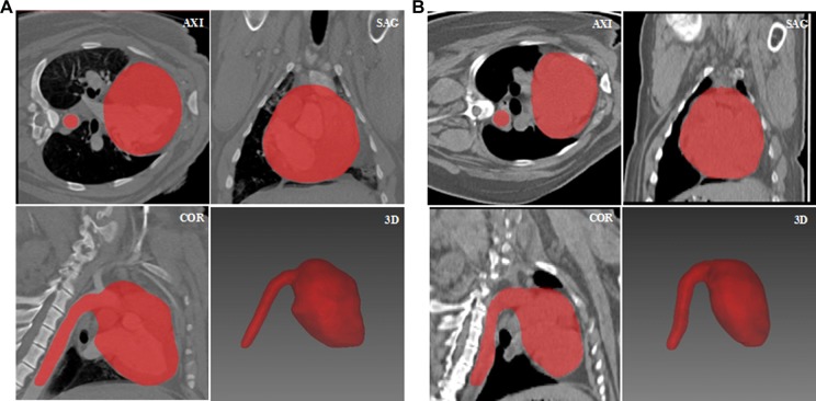
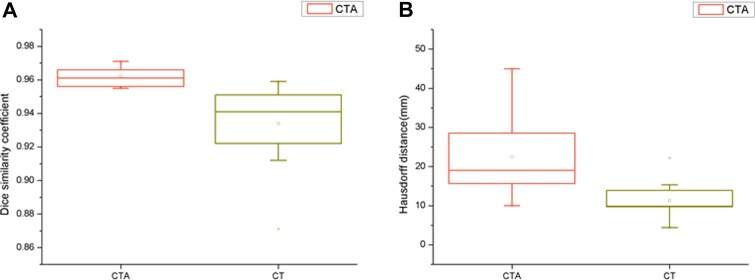
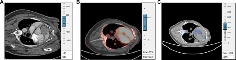
The combination of different modality images can provide detailed and comprehensive information for the prognostic assessment and therapeutic strategy of patients with ischemic heart disease. In this study, a 3D fusion framework is designed to integrate coronary computed tomography (CT) angiography (CTA), 2-deoxy-2-[18F]fluoro-D-glucose ([18F]DG) positron emission tomography (PET)/CT, and [68Ga]-1,4,7-triazacyclononane-1,4,7-triacetic acid-(Arg-Gly-Asp)2 ([68Ga]-NOTA-PRGD2) PET/CT images of the myocardial infarction model in minipigs. First, the structural anatomy of the heart in coronary CTA and CT is segmented using a multi-atlas-based method. Then, the hearts are registered using the B-spline-based free form deformation. Finally, the [18F]DG and [68Ga]-NOTA-PRGD2 signals are mapped into the heart in coronary CTA, which produces a single fusion image to delineate both the cardiac structural anatomy and the functional information of myocardial viability and angiogenesis. Heart segmentation demonstrates high accuracy with good agreement between manual delineation and automatic segmentation. The fusion result intuitively reflects the extent of the [18F]DG uptake defect as well as the location where the [68Ga]-NOTA-PRGD2 signal appears. The fusion result verified the occurrence of angiogenesis based on the in vivo noninvasive molecular imaging approach. The presented framework is helpful in facilitating the study of the relationship between infarct territories and blocked coronary arteries as well as angiogenesis.
Molecular ImagingBiochemistry, Genetics and Molecular Biology-Biotechnology
自引率
3.60%
发文量
21
期刊介绍:
Molecular Imaging is a peer-reviewed, open access journal highlighting the breadth of molecular imaging research from basic science to preclinical studies to human applications. This serves both the scientific and clinical communities by disseminating novel results and concepts relevant to the biological study of normal and disease processes in both basic and translational studies ranging from mice to humans.


 分享
分享
 求助内容:
求助内容: 应助结果提醒方式:
 扫码关注我们
