Hui Liu, Hongjun Jin, Xuyi Yue, Junbin Han, Pamela Baum, Dana R Abendschein, Zhude Tu
{"title":"PET Study of Sphingosine-1-Phosphate Receptor 1 Expression in Response to Vascular Inflammation in a Rat Model of Carotid Injury.","authors":"Hui Liu, Hongjun Jin, Xuyi Yue, Junbin Han, Pamela Baum, Dana R Abendschein, Zhude Tu","doi":"10.1177/1536012116689770","DOIUrl":null,"url":null,"abstract":"<p><p>Sphingosine-1-phosphate receptor (S1PR) activation plays a key role in vascular inflammatory response. Here, we report in vivo validation of [<sup>11</sup>C]TZ3321, a potent S1PR1 radioligand, for imaging vascular inflammation in a rat model of carotid injury. The right common carotid artery of male adult Sprague-Dawley rats was injured by balloon overinflation that denuded the endothelium and distended the vessel wall. Animals received a 60-minute micro-positron emission tomography (micro PET) scan with [<sup>11</sup>C]TZ3321 at 72 hours after injury. Ex vivo autoradiography was also conducted. The expression and cellular location of S1PR1 were examined by immunohistological analysis. Three-dimensional (3D) reconstruction of the first 100-second microPET/computed tomography (CT) image indicated the location of bilateral common carotid arteries. [<sup>11</sup>C]TZ3321 displayed significantly higher accumulation (standardized uptake values: 0.93 ± 0.07 vs 0.78 ± 0.09, n = 6, P = .001) in the injured carotid artery than in the contralateral side. Increased tracer uptake in the injured artery was confirmed by autoradiography (photostimulated luminescence measures: 85.5 ± 0.93 vs 71.48 ± 6.22, n = 2). Concordantly, high S1PR1expression was observed in infiltrated inflammatory cells in the injured artery. Our studies demonstrate [<sup>11</sup>C]TZ3321 microPET is able to detect the acute upregulation of S1PR1 expression in inflamed carotid artery. Therefore, [<sup>11</sup>C]TZ3321 has potential to be a PET radiotracer for detecting early inflammatory response and monitoring therapeutic efficacy of vascular inflammation.</p>","PeriodicalId":18855,"journal":{"name":"Molecular Imaging","volume":"16 ","pages":"1536012116689770"},"PeriodicalIF":2.4000,"publicationDate":"2017-01-01","publicationTypes":"Journal Article","fieldsOfStudy":null,"isOpenAccess":false,"openAccessPdf":"https://sci-hub-pdf.com/10.1177/1536012116689770","citationCount":"19","resultStr":null,"platform":"Semanticscholar","paperid":null,"PeriodicalName":"Molecular Imaging","FirstCategoryId":"3","ListUrlMain":"https://doi.org/10.1177/1536012116689770","RegionNum":4,"RegionCategory":"医学","ArticlePicture":[],"TitleCN":null,"AbstractTextCN":null,"PMCID":null,"EPubDate":"","PubModel":"","JCR":"Q3","JCRName":"BIOCHEMICAL RESEARCH METHODS","Score":null,"Total":0}
引用次数: 19
Abstract
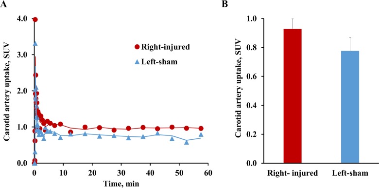
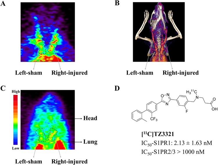
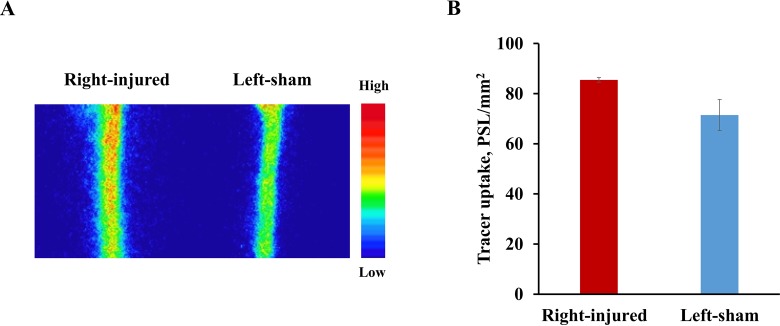
Sphingosine-1-phosphate receptor (S1PR) activation plays a key role in vascular inflammatory response. Here, we report in vivo validation of [11C]TZ3321, a potent S1PR1 radioligand, for imaging vascular inflammation in a rat model of carotid injury. The right common carotid artery of male adult Sprague-Dawley rats was injured by balloon overinflation that denuded the endothelium and distended the vessel wall. Animals received a 60-minute micro-positron emission tomography (micro PET) scan with [11C]TZ3321 at 72 hours after injury. Ex vivo autoradiography was also conducted. The expression and cellular location of S1PR1 were examined by immunohistological analysis. Three-dimensional (3D) reconstruction of the first 100-second microPET/computed tomography (CT) image indicated the location of bilateral common carotid arteries. [11C]TZ3321 displayed significantly higher accumulation (standardized uptake values: 0.93 ± 0.07 vs 0.78 ± 0.09, n = 6, P = .001) in the injured carotid artery than in the contralateral side. Increased tracer uptake in the injured artery was confirmed by autoradiography (photostimulated luminescence measures: 85.5 ± 0.93 vs 71.48 ± 6.22, n = 2). Concordantly, high S1PR1expression was observed in infiltrated inflammatory cells in the injured artery. Our studies demonstrate [11C]TZ3321 microPET is able to detect the acute upregulation of S1PR1 expression in inflamed carotid artery. Therefore, [11C]TZ3321 has potential to be a PET radiotracer for detecting early inflammatory response and monitoring therapeutic efficacy of vascular inflammation.
鞘氨醇-1-磷酸受体(S1PR)的激活在血管炎症反应中起关键作用。在这里,我们报道了[11C]TZ3321(一种有效的S1PR1放射配体)在大鼠颈动脉损伤模型中用于血管炎症成像的体内验证。雄性成年sd大鼠右颈总动脉球囊过度膨胀,内皮脱落,血管壁扩张。动物在损伤后72小时用[11C]TZ3321进行60分钟的微正电子发射断层扫描(micro PET)。还进行了离体放射自显影。免疫组织学分析检测S1PR1的表达及细胞定位。首100秒微pet /计算机断层扫描(CT)图像的三维重建显示了双侧颈总动脉的位置。[11C]TZ3321在损伤颈动脉中的累积量(标准化摄取值:0.93±0.07 vs 0.78±0.09,n = 6, P = 0.001)明显高于对侧。放射自显影证实损伤动脉示踪剂摄取增加(光刺激发光测量:85.5±0.93 vs 71.48±6.22,n = 2)。同时,在损伤动脉浸润炎性细胞中观察到高s1pr1表达。我们的研究表明[11C]TZ3321微pet能够检测炎症颈动脉中S1PR1表达的急性上调。因此,[11C]TZ3321有潜力成为PET放射性示踪剂,用于检测早期炎症反应和监测血管炎症的治疗效果。
Molecular ImagingBiochemistry, Genetics and Molecular Biology-Biotechnology
自引率
3.60%
发文量
21
期刊介绍:
Molecular Imaging is a peer-reviewed, open access journal highlighting the breadth of molecular imaging research from basic science to preclinical studies to human applications. This serves both the scientific and clinical communities by disseminating novel results and concepts relevant to the biological study of normal and disease processes in both basic and translational studies ranging from mice to humans.


 分享
分享
 求助内容:
求助内容: 应助结果提醒方式:
 扫码关注我们
