Multimodal Imaging of Patients With Gliomas Confirms 11C-MET PET as a Complementary Marker to MRI for Noninvasive Tumor Grading and Intraindividual Follow-Up After Therapy.
Kai R Laukamp, Florian Lindemann, Matthias Weckesser, Volker Hesselmann, Sandra Ligges, Johannes Wölfer, Astrid Jeibmann, Bastian Zinnhardt, Thomas Viel, Michael Schäfers, Werner Paulus, Walter Stummer, Otmar Schober, Andreas H Jacobs
{"title":"Multimodal Imaging of Patients With Gliomas Confirms <sup>11</sup>C-MET PET as a Complementary Marker to MRI for Noninvasive Tumor Grading and Intraindividual Follow-Up After Therapy.","authors":"Kai R Laukamp, Florian Lindemann, Matthias Weckesser, Volker Hesselmann, Sandra Ligges, Johannes Wölfer, Astrid Jeibmann, Bastian Zinnhardt, Thomas Viel, Michael Schäfers, Werner Paulus, Walter Stummer, Otmar Schober, Andreas H Jacobs","doi":"10.1177/1536012116687651","DOIUrl":null,"url":null,"abstract":"<p><p>The value of combined L-( methyl-[<sup>11</sup>C]) methionine positron-emitting tomography (MET-PET) and magnetic resonance imaging (MRI) with regard to tumor extent, entity prediction, and therapy effects in clinical routine in patients with suspicion of a brain tumor was investigated. In n = 65 patients with histologically verified brain lesions n = 70 MET-PET and MRI (T1-weighted gadolinium-enhanced [T1w-Gd] and fluid-attenuated inversion recovery or T2-weighted [FLAIR/T2w]) examinations were performed. The computer software \"visualization and analysis framework volume rendering engine (Voreen)\" was used for analysis of extent and intersection of tumor compartments. Binary logistic regression models were developed to differentiate between World Health Organization (WHO) tumor types/grades. Tumor sizes as defined by thresholding based on tumor-to-background ratios were significantly different as determined by MET-PET (21.6 ± 36.8 cm<sup>3</sup>), T1w-Gd-MRI (3.9 ± 7.8 cm<sup>3</sup>), and FLAIR/T2-MRI (64.8 ± 60.4 cm<sup>3</sup>; P < .001). The MET-PET visualized tumor activity where MRI parameters were negative: PET positive tumor volume without Gd enhancement was 19.8 ± 35.0 cm<sup>3</sup> and without changes in FLAIR/T2 10.3 ± 25.7 cm<sup>3</sup>. FLAIR/T2-MRI visualized greatest tumor extent with differences to MET-PET being greater in posttherapy (64.6 ± 62.7 cm<sup>3</sup>) than in newly diagnosed patients (20.5 ± 52.6 cm<sup>3</sup>). The binary logistic regression model differentiated between WHO tumor types (fibrillary astrocytoma II n = 10 from other gliomas n = 16) with an accuracy of 80.8% in patients at primary diagnosis. Combined PET and MRI improve the evaluation of tumor activity, extent, type/grade prediction, and therapy-induced changes in patients with glioma and serve information highly relevant for diagnosis and management.</p>","PeriodicalId":18855,"journal":{"name":"Molecular Imaging","volume":"16 ","pages":"1536012116687651"},"PeriodicalIF":2.4000,"publicationDate":"2017-01-01","publicationTypes":"Journal Article","fieldsOfStudy":null,"isOpenAccess":false,"openAccessPdf":"https://sci-hub-pdf.com/10.1177/1536012116687651","citationCount":"18","resultStr":null,"platform":"Semanticscholar","paperid":null,"PeriodicalName":"Molecular Imaging","FirstCategoryId":"3","ListUrlMain":"https://doi.org/10.1177/1536012116687651","RegionNum":4,"RegionCategory":"医学","ArticlePicture":[],"TitleCN":null,"AbstractTextCN":null,"PMCID":null,"EPubDate":"","PubModel":"","JCR":"Q3","JCRName":"BIOCHEMICAL RESEARCH METHODS","Score":null,"Total":0}
引用次数: 18
Abstract
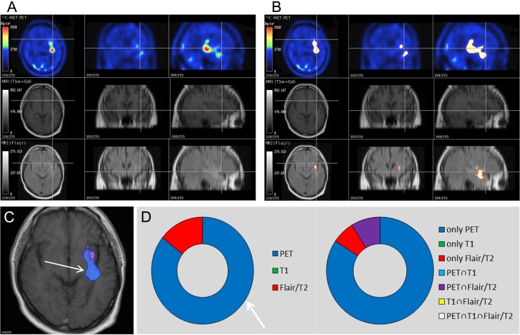
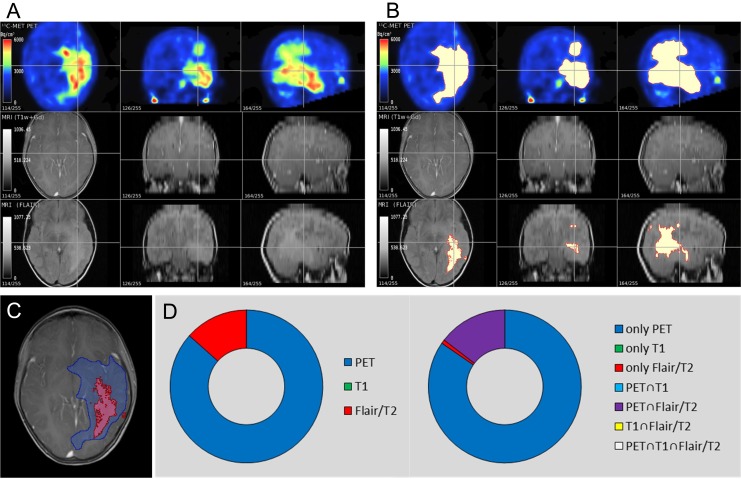
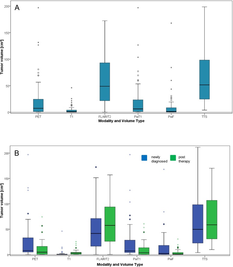
The value of combined L-( methyl-[11C]) methionine positron-emitting tomography (MET-PET) and magnetic resonance imaging (MRI) with regard to tumor extent, entity prediction, and therapy effects in clinical routine in patients with suspicion of a brain tumor was investigated. In n = 65 patients with histologically verified brain lesions n = 70 MET-PET and MRI (T1-weighted gadolinium-enhanced [T1w-Gd] and fluid-attenuated inversion recovery or T2-weighted [FLAIR/T2w]) examinations were performed. The computer software "visualization and analysis framework volume rendering engine (Voreen)" was used for analysis of extent and intersection of tumor compartments. Binary logistic regression models were developed to differentiate between World Health Organization (WHO) tumor types/grades. Tumor sizes as defined by thresholding based on tumor-to-background ratios were significantly different as determined by MET-PET (21.6 ± 36.8 cm3), T1w-Gd-MRI (3.9 ± 7.8 cm3), and FLAIR/T2-MRI (64.8 ± 60.4 cm3; P < .001). The MET-PET visualized tumor activity where MRI parameters were negative: PET positive tumor volume without Gd enhancement was 19.8 ± 35.0 cm3 and without changes in FLAIR/T2 10.3 ± 25.7 cm3. FLAIR/T2-MRI visualized greatest tumor extent with differences to MET-PET being greater in posttherapy (64.6 ± 62.7 cm3) than in newly diagnosed patients (20.5 ± 52.6 cm3). The binary logistic regression model differentiated between WHO tumor types (fibrillary astrocytoma II n = 10 from other gliomas n = 16) with an accuracy of 80.8% in patients at primary diagnosis. Combined PET and MRI improve the evaluation of tumor activity, extent, type/grade prediction, and therapy-induced changes in patients with glioma and serve information highly relevant for diagnosis and management.
Molecular ImagingBiochemistry, Genetics and Molecular Biology-Biotechnology
自引率
3.60%
发文量
21
期刊介绍:
Molecular Imaging is a peer-reviewed, open access journal highlighting the breadth of molecular imaging research from basic science to preclinical studies to human applications. This serves both the scientific and clinical communities by disseminating novel results and concepts relevant to the biological study of normal and disease processes in both basic and translational studies ranging from mice to humans.


 分享
分享
 求助内容:
求助内容: 应助结果提醒方式:
 扫码关注我们
