Benyamin Rosental, Zhanna Kozhekbaeva, Nathaniel Fernhoff, Jonathan M Tsai, Nikki Traylor-Knowles
{"title":"Coral cell separation and isolation by fluorescence-activated cell sorting (FACS).","authors":"Benyamin Rosental, Zhanna Kozhekbaeva, Nathaniel Fernhoff, Jonathan M Tsai, Nikki Traylor-Knowles","doi":"10.1186/s12860-017-0146-8","DOIUrl":null,"url":null,"abstract":"<p><strong>Background: </strong>Generalized methods for understanding the cell biology of non-model species are quite rare, yet very much needed. In order to address this issue, we have modified a technique traditionally used in the biomedical field for ecological and evolutionary research. Fluorescent activated cell sorting (FACS) is often used for sorting and identifying cell populations. In this study, we developed a method to identify and isolate different cell populations in corals and other cnidarians.</p><p><strong>Methods: </strong>Using fluorescence-activated cell sorting (FACS), coral cell suspension were sorted into different cellular populations using fluorescent cell markers that are non-species specific. Over 30 different cell markers were tested. Additionally, cell suspension from Aiptasia pallida was also tested, and a phagocytosis test was done as a downstream functional assay.</p><p><strong>Results: </strong>We found that 24 of the screened markers positively labeled coral cells and 16 differentiated cell sub-populations. We identified 12 different cellular sub-populations using three markers, and found that each sub-population is primarily homogeneous. Lastly, we verified this technique in a sea anemone, Aiptasia pallida, and found that with minor modifications, a similar gating strategy can be successfully applied. Additionally, within A. pallida, we show elevated phagocytosis of sorted cells based on an immune associated marker.</p><p><strong>Conclusions: </strong>In this study, we successfully adapted FACS for isolating coral cell populations and conclude that this technique is translatable for future use in other species. This technique has the potential to be used for different types of studies on the cellular stress response and other immunological studies.</p>","PeriodicalId":9051,"journal":{"name":"BMC Cell Biology","volume":"18 1","pages":"30"},"PeriodicalIF":0.0000,"publicationDate":"2017-08-29","publicationTypes":"Journal Article","fieldsOfStudy":null,"isOpenAccess":false,"openAccessPdf":"https://www.ncbi.nlm.nih.gov/pmc/articles/PMC5575905/pdf/","citationCount":"0","resultStr":null,"platform":"Semanticscholar","paperid":null,"PeriodicalName":"BMC Cell Biology","FirstCategoryId":"1085","ListUrlMain":"https://doi.org/10.1186/s12860-017-0146-8","RegionNum":0,"RegionCategory":null,"ArticlePicture":[],"TitleCN":null,"AbstractTextCN":null,"PMCID":null,"EPubDate":"","PubModel":"","JCR":"Q1","JCRName":"Biochemistry, Genetics and Molecular Biology","Score":null,"Total":0}
引用次数: 0
Abstract
Background: Generalized methods for understanding the cell biology of non-model species are quite rare, yet very much needed. In order to address this issue, we have modified a technique traditionally used in the biomedical field for ecological and evolutionary research. Fluorescent activated cell sorting (FACS) is often used for sorting and identifying cell populations. In this study, we developed a method to identify and isolate different cell populations in corals and other cnidarians.
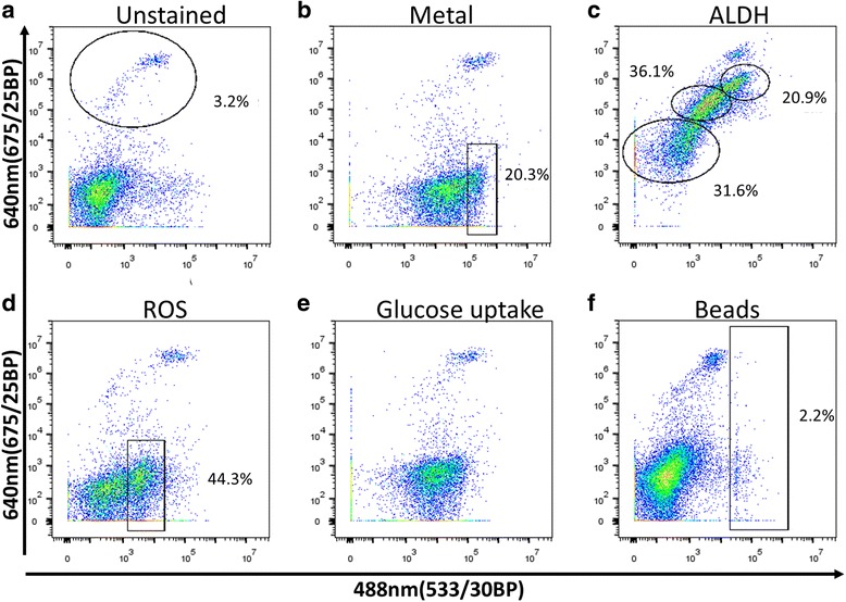
Methods: Using fluorescence-activated cell sorting (FACS), coral cell suspension were sorted into different cellular populations using fluorescent cell markers that are non-species specific. Over 30 different cell markers were tested. Additionally, cell suspension from Aiptasia pallida was also tested, and a phagocytosis test was done as a downstream functional assay.
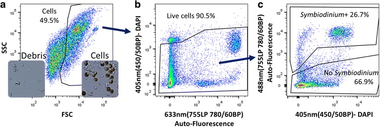
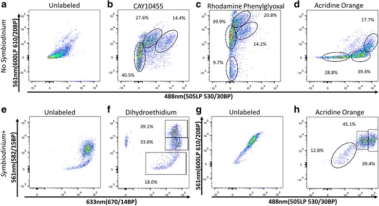
Results: We found that 24 of the screened markers positively labeled coral cells and 16 differentiated cell sub-populations. We identified 12 different cellular sub-populations using three markers, and found that each sub-population is primarily homogeneous. Lastly, we verified this technique in a sea anemone, Aiptasia pallida, and found that with minor modifications, a similar gating strategy can be successfully applied. Additionally, within A. pallida, we show elevated phagocytosis of sorted cells based on an immune associated marker.
Conclusions: In this study, we successfully adapted FACS for isolating coral cell populations and conclude that this technique is translatable for future use in other species. This technique has the potential to be used for different types of studies on the cellular stress response and other immunological studies.
期刊介绍:
BMC Molecular and Cell Biology, formerly known as BMC Cell Biology, is an open access journal that considers articles on all aspects of both eukaryotic and prokaryotic cell and molecular biology, including structural and functional cell biology, DNA and RNA in a cellular context and biochemistry, as well as research using both the experimental and theoretical aspects of physics to study biological processes and investigations into the structure of biological macromolecules.


 分享
分享
 求助内容:
求助内容: 应助结果提醒方式:
 扫码关注我们
