Hakan Bilhan, Onur Geckili, Selda Arat Bilhan, Fatih Aycicek, Berkman Albayrak, Pelin Bozbulut, Fatma Unalan
{"title":"The comparison of the precision of different dental radiographic methods in mandibular peri-implantary measurements: an in vitro study.","authors":"Hakan Bilhan, Onur Geckili, Selda Arat Bilhan, Fatih Aycicek, Berkman Albayrak, Pelin Bozbulut, Fatma Unalan","doi":"10.17096/jiufd.55134","DOIUrl":null,"url":null,"abstract":"<p><strong>Purpose: </strong>The objective of this in vitro study was to investigate and compare the precisions of several radiodiagnostic methods used in dentistry for the measurement of peri-implantary sites.</p><p><strong>Materials and methods: </strong>Six dental implants were placed in a human cadaver mandible. Periapical radiographs obtained with the parallel as well as the bisecting angle technique, digital and conventional panoramic radiographs were used for implant and peri-implant bone measurements. The measurement results at each implant were statistically analyzed.</p><p><strong>Results: </strong>The ICC values for the inter-observer reliability were 0.79 for implant diameters and 0.96 for implant lengths. Statistical significance was not detected between the differences of the measurements of the 2 examiners from the original implant dimensions related to anatomic locations. For both of the examiner measurements, significantly less difference from the original implant dimensions was detected in the parallel technique compared to the other techniques (p<0.05).</p><p><strong>Conclusion: </strong>The present study showed that the most precise peri-implant bone measurements can be obtained from periapical radiographies by using the parallel technique.</p>","PeriodicalId":30947,"journal":{"name":"Journal of Istanbul University Faculty of Dentistry","volume":"49 1","pages":"1-9"},"PeriodicalIF":0.0000,"publicationDate":"2015-01-31","publicationTypes":"Journal Article","fieldsOfStudy":null,"isOpenAccess":false,"openAccessPdf":"https://ftp.ncbi.nlm.nih.gov/pub/pmc/oa_pdf/a3/5a/jiufd-049-001-a.PMC5573457.pdf","citationCount":"3","resultStr":null,"platform":"Semanticscholar","paperid":null,"PeriodicalName":"Journal of Istanbul University Faculty of Dentistry","FirstCategoryId":"1085","ListUrlMain":"https://doi.org/10.17096/jiufd.55134","RegionNum":0,"RegionCategory":null,"ArticlePicture":[],"TitleCN":null,"AbstractTextCN":null,"PMCID":null,"EPubDate":"2015/1/1 0:00:00","PubModel":"eCollection","JCR":"","JCRName":"","Score":null,"Total":0}
引用次数: 3
Abstract
Purpose: The objective of this in vitro study was to investigate and compare the precisions of several radiodiagnostic methods used in dentistry for the measurement of peri-implantary sites.
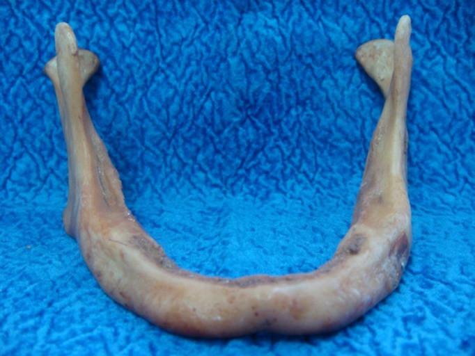
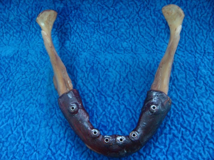
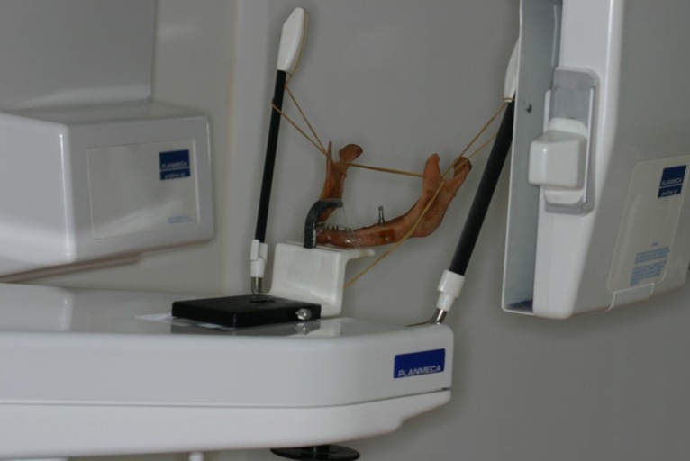
Materials and methods: Six dental implants were placed in a human cadaver mandible. Periapical radiographs obtained with the parallel as well as the bisecting angle technique, digital and conventional panoramic radiographs were used for implant and peri-implant bone measurements. The measurement results at each implant were statistically analyzed.
Results: The ICC values for the inter-observer reliability were 0.79 for implant diameters and 0.96 for implant lengths. Statistical significance was not detected between the differences of the measurements of the 2 examiners from the original implant dimensions related to anatomic locations. For both of the examiner measurements, significantly less difference from the original implant dimensions was detected in the parallel technique compared to the other techniques (p<0.05).
Conclusion: The present study showed that the most precise peri-implant bone measurements can be obtained from periapical radiographies by using the parallel technique.


 分享
分享

 求助内容:
求助内容: 应助结果提醒方式:
 扫码关注我们
