{"title":"Detailed analysis of a superficial CD34-positive fibroblastic tumor: A case report and review of the literature.","authors":"Kensaku Yamaga, Akihiro Fujita, Mari Osaki, Satoshi Kuwamoto, Naoko Ishiguro, Tadahito Yamamoto, Hideki Nagashima","doi":"10.3892/ol.2017.6636","DOIUrl":null,"url":null,"abstract":"<p><p>Superficial cluster of differentiation (CD)34-positive fibroblastic tumor (SCPFT) is a rare mesenchymal neoplasm of borderline malignancy. It is characterized by a superficial location, marked cellular pleomorphism, an extremely low incidence of mitotic figures, and strong CD34 immunohistochemical positivity. As SCPFT is a recently described neoplasm, its characteristics are yet to be fully elucidated. To the best of our knowledge, no detailed studies regarding the imaging findings and cytogenetic analyses of SCPFTs exist. The present study describes a typical case of an 18-year-old man who developed an SCPFT measuring 87×70×80 mm in the subcutaneous adipose tissue of his right thigh. Computed tomography (CT) revealed a well-marginated tumor without calcification, and the enhancement on CT was weak. The tumor demonstrated abnormal uptake on 2-(18F) fluoro-2-deoxy-D-glucose positron emission tomography (PET), with a maximum standardized uptake value of 2.57. Magnetic resonance imaging (MRI) revealed a clearly defined tumor that exhibited homogeneous low signal intensity on T1-weighted imaging and high signal intensity on T2-weighted imaging, with small lobulated structures. Histopathologically, the tumor was composed of irregular spindle-to-oval-shaped cells with eosinophilic glassy cytoplasm and hyperchromatic, bizarre and pleomorphic nuclei that frequently exhibited intranuclear pseudoinclusions. Immunohistochemically, the tumor cells were diffusely and strongly positive for CD34. The Mindbomb E3 ubiquitin protein ligase 1 labeling index was 8.6%. Ultrastructurally, the tumor cells exhibited irregular or convoluted nuclei with abundant euchromatin-prominent nucleoli. The cytoplasmic organelles consisted of scattered, abundant rough endoplasmic reticulum, mitochondria, lysosomes, ribosomal rosettes and aggregated lipid globules. Of 18 metaphase cells identified, 2 demonstrated translocation between chromosomes 2 and 5 in cytogenetic studies. To the best of our knowledge, this is the first study describing imaging data (CT, MRI and PET-CT) and chromosomal aberrations for SCPFT.</p>","PeriodicalId":19503,"journal":{"name":"Oncology Letters","volume":" ","pages":"3395-3400"},"PeriodicalIF":2.2000,"publicationDate":"2017-09-01","publicationTypes":"Journal Article","fieldsOfStudy":null,"isOpenAccess":false,"openAccessPdf":"https://sci-hub-pdf.com/10.3892/ol.2017.6636","citationCount":"17","resultStr":null,"platform":"Semanticscholar","paperid":null,"PeriodicalName":"Oncology Letters","FirstCategoryId":"3","ListUrlMain":"https://doi.org/10.3892/ol.2017.6636","RegionNum":4,"RegionCategory":"医学","ArticlePicture":[],"TitleCN":null,"AbstractTextCN":null,"PMCID":null,"EPubDate":"2017/7/20 0:00:00","PubModel":"Epub","JCR":"Q3","JCRName":"ONCOLOGY","Score":null,"Total":0}
引用次数: 17
Abstract
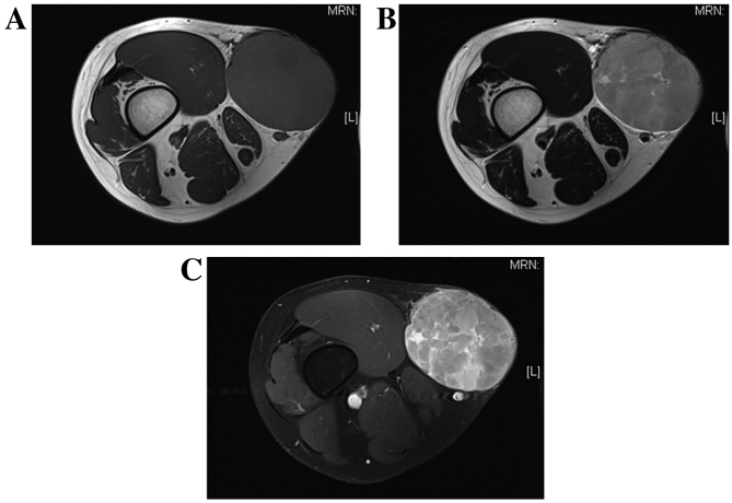
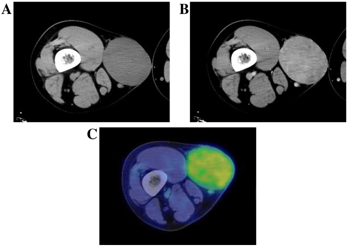
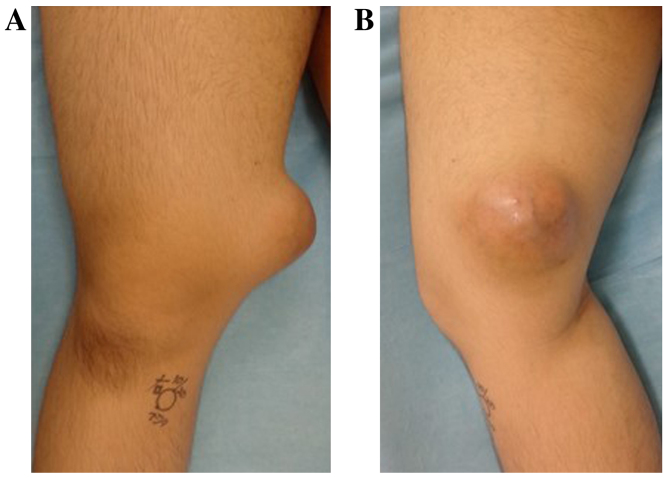
Superficial cluster of differentiation (CD)34-positive fibroblastic tumor (SCPFT) is a rare mesenchymal neoplasm of borderline malignancy. It is characterized by a superficial location, marked cellular pleomorphism, an extremely low incidence of mitotic figures, and strong CD34 immunohistochemical positivity. As SCPFT is a recently described neoplasm, its characteristics are yet to be fully elucidated. To the best of our knowledge, no detailed studies regarding the imaging findings and cytogenetic analyses of SCPFTs exist. The present study describes a typical case of an 18-year-old man who developed an SCPFT measuring 87×70×80 mm in the subcutaneous adipose tissue of his right thigh. Computed tomography (CT) revealed a well-marginated tumor without calcification, and the enhancement on CT was weak. The tumor demonstrated abnormal uptake on 2-(18F) fluoro-2-deoxy-D-glucose positron emission tomography (PET), with a maximum standardized uptake value of 2.57. Magnetic resonance imaging (MRI) revealed a clearly defined tumor that exhibited homogeneous low signal intensity on T1-weighted imaging and high signal intensity on T2-weighted imaging, with small lobulated structures. Histopathologically, the tumor was composed of irregular spindle-to-oval-shaped cells with eosinophilic glassy cytoplasm and hyperchromatic, bizarre and pleomorphic nuclei that frequently exhibited intranuclear pseudoinclusions. Immunohistochemically, the tumor cells were diffusely and strongly positive for CD34. The Mindbomb E3 ubiquitin protein ligase 1 labeling index was 8.6%. Ultrastructurally, the tumor cells exhibited irregular or convoluted nuclei with abundant euchromatin-prominent nucleoli. The cytoplasmic organelles consisted of scattered, abundant rough endoplasmic reticulum, mitochondria, lysosomes, ribosomal rosettes and aggregated lipid globules. Of 18 metaphase cells identified, 2 demonstrated translocation between chromosomes 2 and 5 in cytogenetic studies. To the best of our knowledge, this is the first study describing imaging data (CT, MRI and PET-CT) and chromosomal aberrations for SCPFT.
期刊介绍:
Oncology Letters is a monthly, peer-reviewed journal, available in print and online, that focuses on all aspects of clinical oncology, as well as in vitro and in vivo experimental model systems relevant to the mechanisms of disease.
The principal aim of Oncology Letters is to provide the prompt publication of original studies of high quality that pertain to clinical oncology, chemotherapy, oncogenes, carcinogenesis, metastasis, epidemiology and viral oncology in the form of original research, reviews and case reports.


 分享
分享
 求助内容:
求助内容: 应助结果提醒方式:
 扫码关注我们
