{"title":"In vitro comparison of gutta-percha-filled area percentages in root canals instrumented and obturated with different techniques.","authors":"Ayca Yilmaz, Isil Karagoz-Kucukay","doi":"10.17096/jiufd.95769","DOIUrl":null,"url":null,"abstract":"<p><strong>Purpose: </strong>To evaluate the efficacy of different obturation techniques in root canals instrumented either by hand or rotary instruments with regard to the percentage of gutta- percha-filled area (PGFA).</p><p><strong>Materials and methods: </strong>One hundred and sixty extracted mandibular premolars with single, straight root canals were studied. Root canals were prepared to an apical size of 30 by hand with a modified crown-down technique or the ProTaper and HEROShaper systems. Teeth were divided into eight groups (n=20) according to the following instrumentation and obturation techniques: G1: Hand files+lateral condensation (LC), G2: Hand files+Thermafil, G3: ProTaper+LC, G4: ProTaper+single-cone, G5: ProTaper+ProTaper-Obturator, G6: HEROShaper+LC, G7: HEROShaper+single-cone, G8: HEROShaper+HEROfill. Horizontal sections were cut at 1, 3, 5, 7, 9, 11 and 13 mm from the apical foramen. A total of 1120 sections obtained were digitally photographed under a stereomicroscope set at 48X magnification. The cross-sectional area of the canal and the gutta-percha was measured by digital image analysis and the PGFA was calculated for each section.</p><p><strong>Results: </strong>The mean of the PGFA in Thermafil (G2), ProTaper-Obturator (G5) and HEROfill (G8) groups was significantly higher than the other groups. In G3 and G4, PGFA showed no significant difference in the apical segments whereas PGFA was significantly higher at the middle and coronal segments in G3. In G6 and G7, PGFA showed no significant difference in the apical and middle segments whereas PGFA was significantly higher at the coronal segments in G6.</p><p><strong>Conclusion: </strong>The carrier-based gutta-percha obturation systems revealed significantly higher PGFA in comparison to single-cone and lateral condensation techniques.</p>","PeriodicalId":30947,"journal":{"name":"Journal of Istanbul University Faculty of Dentistry","volume":"51 2","pages":"37-42"},"PeriodicalIF":0.0000,"publicationDate":"2017-04-03","publicationTypes":"Journal Article","fieldsOfStudy":null,"isOpenAccess":false,"openAccessPdf":"https://ftp.ncbi.nlm.nih.gov/pub/pmc/oa_pdf/e4/0f/jiufd-051-037.PMC5573472.pdf","citationCount":"2","resultStr":null,"platform":"Semanticscholar","paperid":null,"PeriodicalName":"Journal of Istanbul University Faculty of Dentistry","FirstCategoryId":"1085","ListUrlMain":"https://doi.org/10.17096/jiufd.95769","RegionNum":0,"RegionCategory":null,"ArticlePicture":[],"TitleCN":null,"AbstractTextCN":null,"PMCID":null,"EPubDate":"2017/1/1 0:00:00","PubModel":"eCollection","JCR":"","JCRName":"","Score":null,"Total":0}
引用次数: 2
Abstract
Purpose: To evaluate the efficacy of different obturation techniques in root canals instrumented either by hand or rotary instruments with regard to the percentage of gutta- percha-filled area (PGFA).
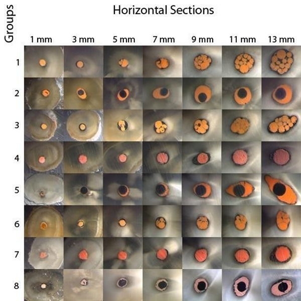
Materials and methods: One hundred and sixty extracted mandibular premolars with single, straight root canals were studied. Root canals were prepared to an apical size of 30 by hand with a modified crown-down technique or the ProTaper and HEROShaper systems. Teeth were divided into eight groups (n=20) according to the following instrumentation and obturation techniques: G1: Hand files+lateral condensation (LC), G2: Hand files+Thermafil, G3: ProTaper+LC, G4: ProTaper+single-cone, G5: ProTaper+ProTaper-Obturator, G6: HEROShaper+LC, G7: HEROShaper+single-cone, G8: HEROShaper+HEROfill. Horizontal sections were cut at 1, 3, 5, 7, 9, 11 and 13 mm from the apical foramen. A total of 1120 sections obtained were digitally photographed under a stereomicroscope set at 48X magnification. The cross-sectional area of the canal and the gutta-percha was measured by digital image analysis and the PGFA was calculated for each section.
Results: The mean of the PGFA in Thermafil (G2), ProTaper-Obturator (G5) and HEROfill (G8) groups was significantly higher than the other groups. In G3 and G4, PGFA showed no significant difference in the apical segments whereas PGFA was significantly higher at the middle and coronal segments in G3. In G6 and G7, PGFA showed no significant difference in the apical and middle segments whereas PGFA was significantly higher at the coronal segments in G6.
Conclusion: The carrier-based gutta-percha obturation systems revealed significantly higher PGFA in comparison to single-cone and lateral condensation techniques.


 分享
分享

 求助内容:
求助内容: 应助结果提醒方式:
 扫码关注我们
