{"title":"Narrow-Energy-Width CT Based on Multivoltage X-Ray Image Decomposition.","authors":"Jiaotong Wei, Yan Han, Ping Chen","doi":"10.1155/2017/8126019","DOIUrl":null,"url":null,"abstract":"<p><p>A polychromatic X-ray beam causes the grey of the reconstructed image to depend on its position within a solid and the material being imaged. This factor makes quantitative measurements via computed tomography (CT) imaging very difficult. To obtain a narrow-energy-width reconstructed image, we propose a model to decompose multivoltage X-ray images into many narrow-energy-width X-ray images by utilizing the low frequency characteristics of X-ray scattering. It needs no change of hardware in the typical CT system. Solving the decomposition model, narrow-energy-width projections are obtained and it is used to reconstruct the image. A cylinder composed of aluminum and silicon is used in a verification experiment. Some of the reconstructed images could be regarded as real narrow-energy-width reconstructed images, which demonstrates the effectiveness of the proposed method.</p>","PeriodicalId":47063,"journal":{"name":"International Journal of Biomedical Imaging","volume":"2017 ","pages":"8126019"},"PeriodicalIF":1.3000,"publicationDate":"2017-01-01","publicationTypes":"Journal Article","fieldsOfStudy":null,"isOpenAccess":false,"openAccessPdf":"https://sci-hub-pdf.com/10.1155/2017/8126019","citationCount":"3","resultStr":null,"platform":"Semanticscholar","paperid":null,"PeriodicalName":"International Journal of Biomedical Imaging","FirstCategoryId":"1085","ListUrlMain":"https://doi.org/10.1155/2017/8126019","RegionNum":0,"RegionCategory":null,"ArticlePicture":[],"TitleCN":null,"AbstractTextCN":null,"PMCID":null,"EPubDate":"2017/11/7 0:00:00","PubModel":"Epub","JCR":"Q2","JCRName":"ENGINEERING, BIOMEDICAL","Score":null,"Total":0}
引用次数: 3
Abstract
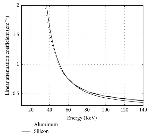
A polychromatic X-ray beam causes the grey of the reconstructed image to depend on its position within a solid and the material being imaged. This factor makes quantitative measurements via computed tomography (CT) imaging very difficult. To obtain a narrow-energy-width reconstructed image, we propose a model to decompose multivoltage X-ray images into many narrow-energy-width X-ray images by utilizing the low frequency characteristics of X-ray scattering. It needs no change of hardware in the typical CT system. Solving the decomposition model, narrow-energy-width projections are obtained and it is used to reconstruct the image. A cylinder composed of aluminum and silicon is used in a verification experiment. Some of the reconstructed images could be regarded as real narrow-energy-width reconstructed images, which demonstrates the effectiveness of the proposed method.
期刊介绍:
The International Journal of Biomedical Imaging is managed by a board of editors comprising internationally renowned active researchers. The journal is freely accessible online and also offered for purchase in print format. It employs a web-based review system to ensure swift turnaround times while maintaining high standards. In addition to regular issues, special issues are organized by guest editors. The subject areas covered include (but are not limited to):
Digital radiography and tomosynthesis
X-ray computed tomography (CT)
Magnetic resonance imaging (MRI)
Single photon emission computed tomography (SPECT)
Positron emission tomography (PET)
Ultrasound imaging
Diffuse optical tomography, coherence, fluorescence, bioluminescence tomography, impedance tomography
Neutron imaging for biomedical applications
Magnetic and optical spectroscopy, and optical biopsy
Optical, electron, scanning tunneling/atomic force microscopy
Small animal imaging
Functional, cellular, and molecular imaging
Imaging assays for screening and molecular analysis
Microarray image analysis and bioinformatics
Emerging biomedical imaging techniques
Imaging modality fusion
Biomedical imaging instrumentation
Biomedical image processing, pattern recognition, and analysis
Biomedical image visualization, compression, transmission, and storage
Imaging and modeling related to systems biology and systems biomedicine
Applied mathematics, applied physics, and chemistry related to biomedical imaging
Grid-enabling technology for biomedical imaging and informatics


 分享
分享
 求助内容:
求助内容: 应助结果提醒方式:
 扫码关注我们
