Alexander James Hale, Ali Kiai, Jelte Sikkens, Jeroen den Hertog
{"title":"Impaired caudal fin-fold regeneration in zebrafish deficient for the tumor suppressor Pten.","authors":"Alexander James Hale, Ali Kiai, Jelte Sikkens, Jeroen den Hertog","doi":"10.1002/reg2.88","DOIUrl":null,"url":null,"abstract":"<p><p>Zebrafish are able to completely regrow their caudal fin-folds after amputation. Following injury, wound healing occurs, followed by the formation of a blastema, which produces cells to replace the lost tissue in the final phase of regenerative outgrowth. Here we show that, surprisingly, the phosphatase and tumor suppressor Pten, an antagonist of phosphoinositide-3-kinase (PI3K) signaling, is required for zebrafish caudal fin-fold regeneration. We found that homozygous knock-out mutant (<i>ptena<sup>-/-</sup>ptenb<sup>-/-</sup></i> ) zebrafish embryos, lacking functional Pten, did not regenerate their caudal fin-folds. AKT phosphorylation was enhanced, which is consistent with the function of Pten. Reexpression of Pten, but not catalytically inactive mutant Pten-C124S, rescued regeneration, as did pharmacological inhibition of PI3K. Blastema formation, determined by in situ hybridization for the blastema marker <i>junbb</i>, appeared normal upon caudal fin-fold amputation of <i>ptena<sup>-/-</sup>ptenb<sup>-/-</sup></i> zebrafish embryos. Whole-mount immunohistochemistry using specific markers indicated that proliferation was arrested in embryos lacking functional Pten, and that apoptosis was enhanced. Together, these results suggest a critical role for Pten by limiting PI3K signaling during the regenerative outgrowth phase of zebrafish caudal fin-fold regeneration.</p>","PeriodicalId":90316,"journal":{"name":"Regeneration (Oxford, England)","volume":"4 4","pages":"217-226"},"PeriodicalIF":0.0000,"publicationDate":"2017-11-10","publicationTypes":"Journal Article","fieldsOfStudy":null,"isOpenAccess":false,"openAccessPdf":"https://www.ncbi.nlm.nih.gov/pmc/articles/PMC5743786/pdf/","citationCount":"0","resultStr":null,"platform":"Semanticscholar","paperid":null,"PeriodicalName":"Regeneration (Oxford, England)","FirstCategoryId":"1085","ListUrlMain":"https://doi.org/10.1002/reg2.88","RegionNum":0,"RegionCategory":null,"ArticlePicture":[],"TitleCN":null,"AbstractTextCN":null,"PMCID":null,"EPubDate":"2017/8/1 0:00:00","PubModel":"eCollection","JCR":"","JCRName":"","Score":null,"Total":0}
引用次数: 0
Abstract
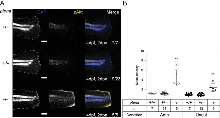
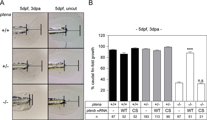
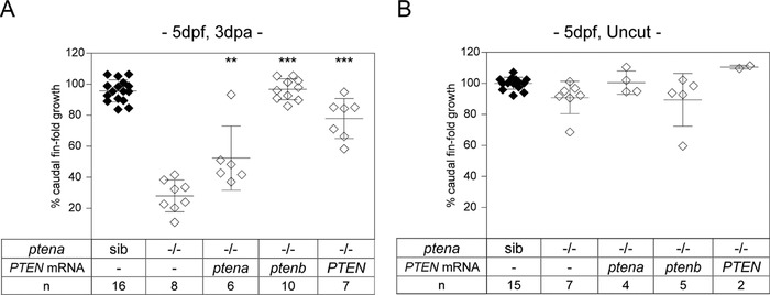
Zebrafish are able to completely regrow their caudal fin-folds after amputation. Following injury, wound healing occurs, followed by the formation of a blastema, which produces cells to replace the lost tissue in the final phase of regenerative outgrowth. Here we show that, surprisingly, the phosphatase and tumor suppressor Pten, an antagonist of phosphoinositide-3-kinase (PI3K) signaling, is required for zebrafish caudal fin-fold regeneration. We found that homozygous knock-out mutant (ptena-/-ptenb-/- ) zebrafish embryos, lacking functional Pten, did not regenerate their caudal fin-folds. AKT phosphorylation was enhanced, which is consistent with the function of Pten. Reexpression of Pten, but not catalytically inactive mutant Pten-C124S, rescued regeneration, as did pharmacological inhibition of PI3K. Blastema formation, determined by in situ hybridization for the blastema marker junbb, appeared normal upon caudal fin-fold amputation of ptena-/-ptenb-/- zebrafish embryos. Whole-mount immunohistochemistry using specific markers indicated that proliferation was arrested in embryos lacking functional Pten, and that apoptosis was enhanced. Together, these results suggest a critical role for Pten by limiting PI3K signaling during the regenerative outgrowth phase of zebrafish caudal fin-fold regeneration.


 分享
分享

 求助内容:
求助内容: 应助结果提醒方式:
 扫码关注我们
