Constantine Zakkaroff, John D Biglands, John P Greenwood, Sven Plein, Roger D Boyle, Aleksandra Radjenovic, Derek R Magee
{"title":"Patient-specific coronary blood supply territories for quantitative perfusion analysis.","authors":"Constantine Zakkaroff, John D Biglands, John P Greenwood, Sven Plein, Roger D Boyle, Aleksandra Radjenovic, Derek R Magee","doi":"10.1080/21681163.2016.1192003","DOIUrl":null,"url":null,"abstract":"<p><p>Myocardial perfusion imaging, coupled with quantitative perfusion analysis, provides an important diagnostic tool for the identification of ischaemic heart disease caused by coronary stenoses. The accurate mapping between coronary anatomy and under-perfused areas of the myocardium is important for diagnosis and treatment. However, in the absence of the actual coronary anatomy during the reporting of perfusion images, areas of ischaemia are allocated to a coronary territory based on a population-derived 17-segment (American Heart Association) AHA model of coronary blood supply. This work presents a solution for the fusion of 2D Magnetic Resonance (MR) myocardial perfusion images and 3D MR angiography data with the aim to improve the detection of ischaemic heart disease. The key contribution of this work is a novel method for the mediated spatiotemporal registration of perfusion and angiography data and a novel method for the calculation of patient-specific coronary supply territories. The registration method uses 4D cardiac MR cine series spanning the complete cardiac cycle in order to overcome the under-constrained nature of non-rigid slice-to-volume perfusion-to-angiography registration. This is achieved by separating out the deformable registration problem and solving it through phase-to-phase registration of the cine series. The use of patient-specific blood supply territories in quantitative perfusion analysis (instead of the population-based model of coronary blood supply) has the potential of increasing the accuracy of perfusion analysis. Quantitative perfusion analysis diagnostic accuracy evaluation with patient-specific territories against the AHA model demonstrates the value of the mediated spatiotemporal registration in the context of ischaemic heart disease diagnosis.</p>","PeriodicalId":51800,"journal":{"name":"Computer Methods in Biomechanics and Biomedical Engineering-Imaging and Visualization","volume":"6 2","pages":"137-154"},"PeriodicalIF":1.3000,"publicationDate":"2016-07-13","publicationTypes":"Journal Article","fieldsOfStudy":null,"isOpenAccess":false,"openAccessPdf":"https://www.ncbi.nlm.nih.gov/pmc/articles/PMC5774224/pdf/","citationCount":"0","resultStr":null,"platform":"Semanticscholar","paperid":null,"PeriodicalName":"Computer Methods in Biomechanics and Biomedical Engineering-Imaging and Visualization","FirstCategoryId":"1085","ListUrlMain":"https://doi.org/10.1080/21681163.2016.1192003","RegionNum":0,"RegionCategory":null,"ArticlePicture":[],"TitleCN":null,"AbstractTextCN":null,"PMCID":null,"EPubDate":"2018/1/1 0:00:00","PubModel":"eCollection","JCR":"Q4","JCRName":"ENGINEERING, BIOMEDICAL","Score":null,"Total":0}
引用次数: 0
Abstract
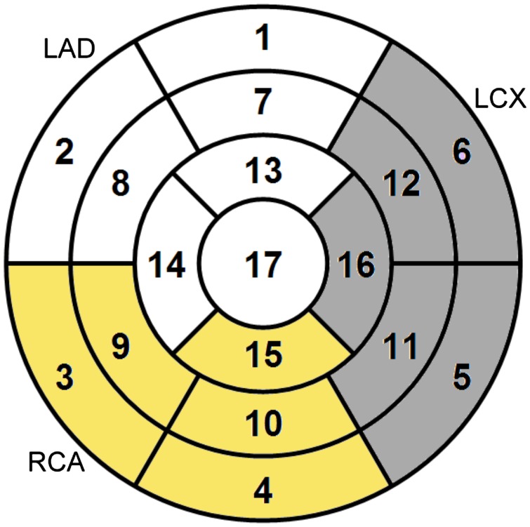
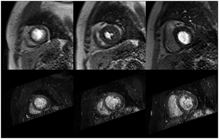
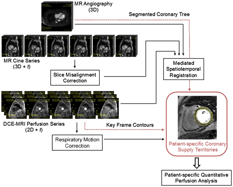
Myocardial perfusion imaging, coupled with quantitative perfusion analysis, provides an important diagnostic tool for the identification of ischaemic heart disease caused by coronary stenoses. The accurate mapping between coronary anatomy and under-perfused areas of the myocardium is important for diagnosis and treatment. However, in the absence of the actual coronary anatomy during the reporting of perfusion images, areas of ischaemia are allocated to a coronary territory based on a population-derived 17-segment (American Heart Association) AHA model of coronary blood supply. This work presents a solution for the fusion of 2D Magnetic Resonance (MR) myocardial perfusion images and 3D MR angiography data with the aim to improve the detection of ischaemic heart disease. The key contribution of this work is a novel method for the mediated spatiotemporal registration of perfusion and angiography data and a novel method for the calculation of patient-specific coronary supply territories. The registration method uses 4D cardiac MR cine series spanning the complete cardiac cycle in order to overcome the under-constrained nature of non-rigid slice-to-volume perfusion-to-angiography registration. This is achieved by separating out the deformable registration problem and solving it through phase-to-phase registration of the cine series. The use of patient-specific blood supply territories in quantitative perfusion analysis (instead of the population-based model of coronary blood supply) has the potential of increasing the accuracy of perfusion analysis. Quantitative perfusion analysis diagnostic accuracy evaluation with patient-specific territories against the AHA model demonstrates the value of the mediated spatiotemporal registration in the context of ischaemic heart disease diagnosis.
期刊介绍:
Computer Methods in Biomechanics and Biomedical Engineering: Imaging & Visualization is an international journal whose main goals are to promote solutions of excellence for both imaging and visualization of biomedical data, and establish links among researchers, clinicians, the medical technology sector and end-users. The journal provides a comprehensive forum for discussion of the current state-of-the-art in the scientific fields related to imaging and visualization, including, but not limited to: Applications of Imaging and Visualization Computational Bio- imaging and Visualization Computer Aided Diagnosis, Surgery, Therapy and Treatment Data Processing and Analysis Devices for Imaging and Visualization Grid and High Performance Computing for Imaging and Visualization Human Perception in Imaging and Visualization Image Processing and Analysis Image-based Geometric Modelling Imaging and Visualization in Biomechanics Imaging and Visualization in Biomedical Engineering Medical Clinics Medical Imaging and Visualization Multi-modal Imaging and Visualization Multiscale Imaging and Visualization Scientific Visualization Software Development for Imaging and Visualization Telemedicine Systems and Applications Virtual Reality Visual Data Mining and Knowledge Discovery.


 分享
分享
 求助内容:
求助内容: 应助结果提醒方式:
 扫码关注我们
