Maria Mihaela Pop, Silviu Cristian, Orsolya Hanko-Bauer, Dana Valentina Ghiga, Rares Georgescu
{"title":"Obtaining adequate surgical margin status in breast-conservation therapy: intraoperative ultrasound-guided resection versus specimen mammography.","authors":"Maria Mihaela Pop, Silviu Cristian, Orsolya Hanko-Bauer, Dana Valentina Ghiga, Rares Georgescu","doi":"10.15386/cjmed-891","DOIUrl":null,"url":null,"abstract":"<p><strong>Background and aim: </strong>The purpose of breast-conserving surgery (BCS) for women with cancer is to perform an oncological radical procedure with disease-free margins at the final histological assessment and with the best aesthetic result possible. Intraoperative resected specimen ultrasound and intraoperative resected specimen mammography may reduce the rates of positive margins and reexcision among patients undergoing conserving therapy. Our objective is to compare the two methods with the histopathological results for a preset cut off and asses which parameters can influence the positive margin status.</p><p><strong>Method: </strong>A prospective study was performed on 83 patients who underwent breast conservation surgery for early breast cancer (pT1-3a pN0-1 M0) between 2014 and 2016. After excision the specimen was oriented in the operating room by the surgeon. Metallic clips and threads were placed on margins: one clip and the long thread at 12 o'clock, two clips and the short threads at 9 o'clock. The next step was intraoperative ultrasound assessment of the specimen. For the margins under 2 mm we performed selective margin shaving, followed by mammography to identify and document the lesion and finally histopathological examination of the specimen with reporting the gross and microscopic margins. The positive margins required re-excision or boost of radiation at the posterior or anterior margins, depending on the case.</p><p><strong>Results: </strong>We set a cut-off at 2 mm. The sensitivity and specificity of the intraoperative margin assessment via the ultrasound method were 90.91% (95% CI 70.84-98.88%) and 67.21% (95% CI 54-78.69%) respectively. The sensitivity and specificity of the intraoperative margin assessment via the mammographic procedure were 45.45% (95% CI 24.39-67.79%) and 85.25% (95% CI 73.83-93.02%) respectively. There was positive correlation between the histopathological and intraoperative ultrasound exam (p=0.018) and negative correlation between the histopathological exam and the post-operative mammographic exam (p=0.68). We found a positive correlation between the positive margin status and age (<40), preoperative chemotherapy, intraductal carcinoma, inflammatory process around the tumor, and the immunohistochemical triple negative profile.</p><p><strong>Conclusions: </strong>According to our results, the intraoperative ultrasound of the breast specimen for a cutt-off at 2 mm can decrease the rates of margin positivity compared to the mammographic procedure and has the potential to diminish the number of subsequent undesired re-excisions.</p>","PeriodicalId":91233,"journal":{"name":"Clujul medical (1957)","volume":"91 2","pages":"197-202"},"PeriodicalIF":0.0000,"publicationDate":"2018-01-01","publicationTypes":"Journal Article","fieldsOfStudy":null,"isOpenAccess":false,"openAccessPdf":"https://sci-hub-pdf.com/10.15386/cjmed-891","citationCount":"9","resultStr":null,"platform":"Semanticscholar","paperid":null,"PeriodicalName":"Clujul medical (1957)","FirstCategoryId":"1085","ListUrlMain":"https://doi.org/10.15386/cjmed-891","RegionNum":0,"RegionCategory":null,"ArticlePicture":[],"TitleCN":null,"AbstractTextCN":null,"PMCID":null,"EPubDate":"2018/4/25 0:00:00","PubModel":"Epub","JCR":"","JCRName":"","Score":null,"Total":0}
引用次数: 9
Abstract
Background and aim: The purpose of breast-conserving surgery (BCS) for women with cancer is to perform an oncological radical procedure with disease-free margins at the final histological assessment and with the best aesthetic result possible. Intraoperative resected specimen ultrasound and intraoperative resected specimen mammography may reduce the rates of positive margins and reexcision among patients undergoing conserving therapy. Our objective is to compare the two methods with the histopathological results for a preset cut off and asses which parameters can influence the positive margin status.
Method: A prospective study was performed on 83 patients who underwent breast conservation surgery for early breast cancer (pT1-3a pN0-1 M0) between 2014 and 2016. After excision the specimen was oriented in the operating room by the surgeon. Metallic clips and threads were placed on margins: one clip and the long thread at 12 o'clock, two clips and the short threads at 9 o'clock. The next step was intraoperative ultrasound assessment of the specimen. For the margins under 2 mm we performed selective margin shaving, followed by mammography to identify and document the lesion and finally histopathological examination of the specimen with reporting the gross and microscopic margins. The positive margins required re-excision or boost of radiation at the posterior or anterior margins, depending on the case.
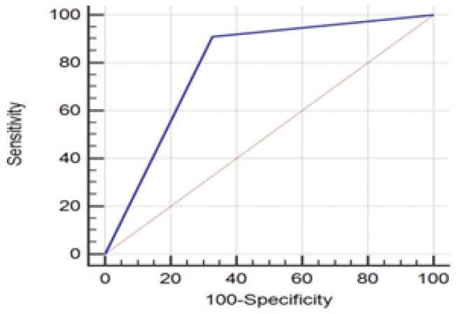
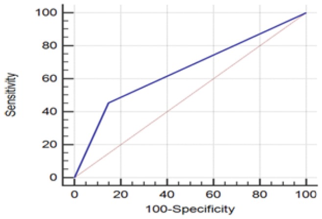
Results: We set a cut-off at 2 mm. The sensitivity and specificity of the intraoperative margin assessment via the ultrasound method were 90.91% (95% CI 70.84-98.88%) and 67.21% (95% CI 54-78.69%) respectively. The sensitivity and specificity of the intraoperative margin assessment via the mammographic procedure were 45.45% (95% CI 24.39-67.79%) and 85.25% (95% CI 73.83-93.02%) respectively. There was positive correlation between the histopathological and intraoperative ultrasound exam (p=0.018) and negative correlation between the histopathological exam and the post-operative mammographic exam (p=0.68). We found a positive correlation between the positive margin status and age (<40), preoperative chemotherapy, intraductal carcinoma, inflammatory process around the tumor, and the immunohistochemical triple negative profile.
Conclusions: According to our results, the intraoperative ultrasound of the breast specimen for a cutt-off at 2 mm can decrease the rates of margin positivity compared to the mammographic procedure and has the potential to diminish the number of subsequent undesired re-excisions.


 分享
分享

 求助内容:
求助内容: 应助结果提醒方式:
 扫码关注我们
