{"title":"Computer-Aided Grading of Gliomas Combining Automatic Segmentation and Radiomics.","authors":"Wei Chen, Boqiang Liu, Suting Peng, Jiawei Sun, Xu Qiao","doi":"10.1155/2018/2512037","DOIUrl":null,"url":null,"abstract":"<p><p>Gliomas are the most common primary brain tumors, and the objective grading is of great importance for treatment. This paper presents an automatic computer-aided diagnosis of gliomas that combines automatic segmentation and radiomics, which can improve the diagnostic ability. The MRI data containing 220 high-grade gliomas and 54 low-grade gliomas are used to evaluate our system. A multiscale 3D convolutional neural network is trained to segment whole tumor regions. A wide range of radiomic features including first-order features, shape features, and texture features is extracted. By using support vector machines with recursive feature elimination for feature selection, a CAD system that has an extreme gradient boosting classifier with a 5-fold cross-validation is constructed for the grading of gliomas. Our CAD system is highly effective for the grading of gliomas with an accuracy of 91.27%, a weighted macroprecision of 91.27%, a weighted macrorecall of 91.27%, and a weighted macro-<i>F</i>1 score of 90.64%. This demonstrates that the proposed CAD system can assist radiologists for high accurate grading of gliomas and has the potential for clinical applications.</p>","PeriodicalId":47063,"journal":{"name":"International Journal of Biomedical Imaging","volume":"2018 ","pages":"2512037"},"PeriodicalIF":1.3000,"publicationDate":"2018-05-08","publicationTypes":"Journal Article","fieldsOfStudy":null,"isOpenAccess":false,"openAccessPdf":"https://sci-hub-pdf.com/10.1155/2018/2512037","citationCount":"60","resultStr":null,"platform":"Semanticscholar","paperid":null,"PeriodicalName":"International Journal of Biomedical Imaging","FirstCategoryId":"1085","ListUrlMain":"https://doi.org/10.1155/2018/2512037","RegionNum":0,"RegionCategory":null,"ArticlePicture":[],"TitleCN":null,"AbstractTextCN":null,"PMCID":null,"EPubDate":"2018/1/1 0:00:00","PubModel":"eCollection","JCR":"Q2","JCRName":"ENGINEERING, BIOMEDICAL","Score":null,"Total":0}
引用次数: 60
Abstract
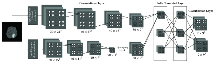
Gliomas are the most common primary brain tumors, and the objective grading is of great importance for treatment. This paper presents an automatic computer-aided diagnosis of gliomas that combines automatic segmentation and radiomics, which can improve the diagnostic ability. The MRI data containing 220 high-grade gliomas and 54 low-grade gliomas are used to evaluate our system. A multiscale 3D convolutional neural network is trained to segment whole tumor regions. A wide range of radiomic features including first-order features, shape features, and texture features is extracted. By using support vector machines with recursive feature elimination for feature selection, a CAD system that has an extreme gradient boosting classifier with a 5-fold cross-validation is constructed for the grading of gliomas. Our CAD system is highly effective for the grading of gliomas with an accuracy of 91.27%, a weighted macroprecision of 91.27%, a weighted macrorecall of 91.27%, and a weighted macro-F1 score of 90.64%. This demonstrates that the proposed CAD system can assist radiologists for high accurate grading of gliomas and has the potential for clinical applications.
期刊介绍:
The International Journal of Biomedical Imaging is managed by a board of editors comprising internationally renowned active researchers. The journal is freely accessible online and also offered for purchase in print format. It employs a web-based review system to ensure swift turnaround times while maintaining high standards. In addition to regular issues, special issues are organized by guest editors. The subject areas covered include (but are not limited to):
Digital radiography and tomosynthesis
X-ray computed tomography (CT)
Magnetic resonance imaging (MRI)
Single photon emission computed tomography (SPECT)
Positron emission tomography (PET)
Ultrasound imaging
Diffuse optical tomography, coherence, fluorescence, bioluminescence tomography, impedance tomography
Neutron imaging for biomedical applications
Magnetic and optical spectroscopy, and optical biopsy
Optical, electron, scanning tunneling/atomic force microscopy
Small animal imaging
Functional, cellular, and molecular imaging
Imaging assays for screening and molecular analysis
Microarray image analysis and bioinformatics
Emerging biomedical imaging techniques
Imaging modality fusion
Biomedical imaging instrumentation
Biomedical image processing, pattern recognition, and analysis
Biomedical image visualization, compression, transmission, and storage
Imaging and modeling related to systems biology and systems biomedicine
Applied mathematics, applied physics, and chemistry related to biomedical imaging
Grid-enabling technology for biomedical imaging and informatics


 分享
分享
 求助内容:
求助内容: 应助结果提醒方式:
 扫码关注我们
