{"title":"Cutaneous dental sinus of submental region: an eight years follow-up.","authors":"Pallav Mahesh Patni, Pradeep Jain, Hemalatha Hiremath, Swadhin Raghuwanshi, Prashansa Vijaywargia, Mona Jain Patni","doi":"10.15386/cjmed-812","DOIUrl":null,"url":null,"abstract":"<p><p>A 22-year-old female patient had a history of a 7-month recurrent pus discharge from her chin. She had been previously treated by physicians, dermatologist, and surgeons. The sinus kept re-occurring and she was referred to dental hospital for opinion. The patient had cutaneous opening of size 5 mm × 6 mm with purulent discharge in submental region. Patient had undergone three surgical excisions and multiple antibiotic regimens. Patient had a history of trauma due to fall six years back. A 30 number standard gutta-percha was used to trace the sinus tract and dental origin was confirmed radiographically. The tract led to in-between the root canal apices of both mandibular incisors. Treatment included non-surgical endodontic treatment with both mandibular central incisors and antibiotic coverage following bacterial culture of discharge. The pus culture showed Streptococcus anginosus which was found to be sensitive to penicillin. Patient was kept on 1-week course of oral amoxicillin-clavulanate along with root canal therapy. The cutaneous sinus healed following root canal treatment and antibiotic coverage. On an 8-year follow-up skin of sub-mental region appeared normal and peri-apical healing with both mandibular central incisors was evident radiographically. Cutaneous lesions on face may be of dental origin. A cross referral between dentists, physicians, surgeons, and dermatologists should be considered in such cases.</p>","PeriodicalId":91233,"journal":{"name":"Clujul medical (1957)","volume":"91 3","pages":"351-356"},"PeriodicalIF":0.0000,"publicationDate":"2018-07-01","publicationTypes":"Journal Article","fieldsOfStudy":null,"isOpenAccess":false,"openAccessPdf":"https://ftp.ncbi.nlm.nih.gov/pub/pmc/oa_pdf/d5/f5/cm-91-351.PMC6082611.pdf","citationCount":"2","resultStr":null,"platform":"Semanticscholar","paperid":null,"PeriodicalName":"Clujul medical (1957)","FirstCategoryId":"1085","ListUrlMain":"https://doi.org/10.15386/cjmed-812","RegionNum":0,"RegionCategory":null,"ArticlePicture":[],"TitleCN":null,"AbstractTextCN":null,"PMCID":null,"EPubDate":"2018/7/31 0:00:00","PubModel":"Epub","JCR":"","JCRName":"","Score":null,"Total":0}
引用次数: 2
Abstract
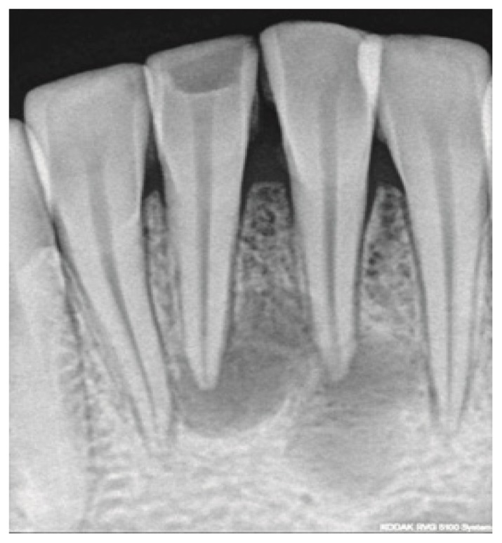
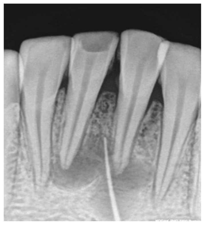
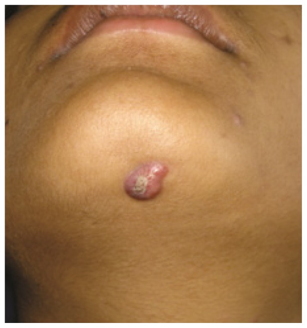
A 22-year-old female patient had a history of a 7-month recurrent pus discharge from her chin. She had been previously treated by physicians, dermatologist, and surgeons. The sinus kept re-occurring and she was referred to dental hospital for opinion. The patient had cutaneous opening of size 5 mm × 6 mm with purulent discharge in submental region. Patient had undergone three surgical excisions and multiple antibiotic regimens. Patient had a history of trauma due to fall six years back. A 30 number standard gutta-percha was used to trace the sinus tract and dental origin was confirmed radiographically. The tract led to in-between the root canal apices of both mandibular incisors. Treatment included non-surgical endodontic treatment with both mandibular central incisors and antibiotic coverage following bacterial culture of discharge. The pus culture showed Streptococcus anginosus which was found to be sensitive to penicillin. Patient was kept on 1-week course of oral amoxicillin-clavulanate along with root canal therapy. The cutaneous sinus healed following root canal treatment and antibiotic coverage. On an 8-year follow-up skin of sub-mental region appeared normal and peri-apical healing with both mandibular central incisors was evident radiographically. Cutaneous lesions on face may be of dental origin. A cross referral between dentists, physicians, surgeons, and dermatologists should be considered in such cases.


 分享
分享

 求助内容:
求助内容: 应助结果提醒方式:
 扫码关注我们
