Can Ultrasound Alone Predict Papillary Thyroid Carcinoma with Desmoid-Type Fibromatosis? A Retrospective Analysis of 13 Cases, Focusing on the Stromal Area.
{"title":"Can Ultrasound Alone Predict Papillary Thyroid Carcinoma with Desmoid-Type Fibromatosis? A Retrospective Analysis of 13 Cases, Focusing on the Stromal Area.","authors":"Kumiko Tajiri, Mitsuyoshi Hirokawa, Ayana Suzuki, Nami Takada, Hisashi Ota, Maki Oshita, Mitsuhiro Fukushima, Kaoru Kobayashi, Akira Miyauchi","doi":"10.1055/a-0591-6163","DOIUrl":null,"url":null,"abstract":"<p><strong>Purpose: </strong>Papillary thyroid carcinoma with desmoid-type fibromatosis (PTC-DTF) is extremely rare. So far, only 4 cases describing the ultrasound findings of this variant have been reported. Here, we describe the ultrasound findings of 13 cases of PTC-DTF, focusing especially on the DTF area.</p><p><strong>Materials and methods: </strong>We retrospectively analyzed the clinical reports, ultrasound reports, and ultrasound photographs obtained from medical records at Kuma Hospital.</p><p><strong>Results: </strong>The patients included 8 women and 5 men with a mean age of 47.9 years. The widest dimension of the nodules ranged from 16 to 79 mm (mean: 37.5 mm). The original ultrasound reports classified the nodules as either intermediate suspicion or high suspicion. A diagnosis of PTC was suspected in 12 nodules, and anaplastic carcinoma was suspected in 1 nodule. PTC-DTF presented with an irregularly shaped nodule (100%), taller-than-wide sign (84.6%), heterogeneous echogenicity (100%), no microcalcification (76.9%), and no or mild flow signal on Doppler (75.0%). The DTF area was identified in the ultrasound photographs of 8 nodules. DTF areas were generally heterogeneous (62.5%) and more hypoechoic (71.4%) than PTC areas. Microcalcification was not observed in the DTF areas. All of the DTF areas revealed no or mild flow signal. On ultrasound elastography, the DTF areas were not stiff, and they were more elastic than the PTC areas.</p><p><strong>Conclusion: </strong>It is difficult to predict PTC-DTF using ultrasound alone, and B-mode ultrasonography is more reliable than ultrasound elastography in the ultrasound diagnosis of malignant thyroid nodules.</p>","PeriodicalId":44852,"journal":{"name":"Ultrasound International Open","volume":"4 2","pages":"E39-E44"},"PeriodicalIF":1.6000,"publicationDate":"2018-04-01","publicationTypes":"Journal Article","fieldsOfStudy":null,"isOpenAccess":false,"openAccessPdf":"https://sci-hub-pdf.com/10.1055/a-0591-6163","citationCount":"7","resultStr":null,"platform":"Semanticscholar","paperid":null,"PeriodicalName":"Ultrasound International Open","FirstCategoryId":"1085","ListUrlMain":"https://doi.org/10.1055/a-0591-6163","RegionNum":0,"RegionCategory":null,"ArticlePicture":[],"TitleCN":null,"AbstractTextCN":null,"PMCID":null,"EPubDate":"2018/7/5 0:00:00","PubModel":"Epub","JCR":"Q3","JCRName":"RADIOLOGY, NUCLEAR MEDICINE & MEDICAL IMAGING","Score":null,"Total":0}
引用次数: 7
Abstract
Purpose: Papillary thyroid carcinoma with desmoid-type fibromatosis (PTC-DTF) is extremely rare. So far, only 4 cases describing the ultrasound findings of this variant have been reported. Here, we describe the ultrasound findings of 13 cases of PTC-DTF, focusing especially on the DTF area.
Materials and methods: We retrospectively analyzed the clinical reports, ultrasound reports, and ultrasound photographs obtained from medical records at Kuma Hospital.
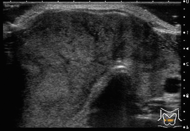
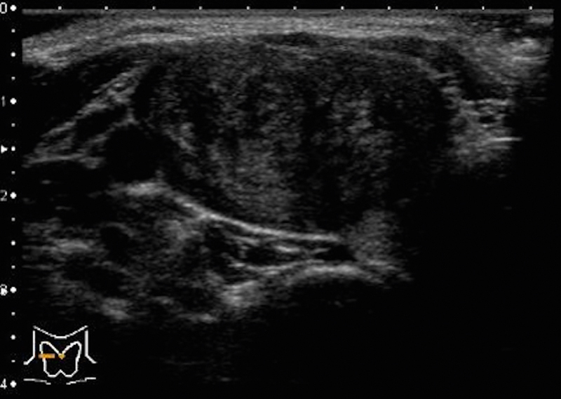
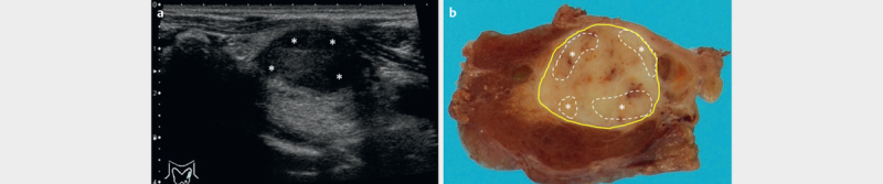
Results: The patients included 8 women and 5 men with a mean age of 47.9 years. The widest dimension of the nodules ranged from 16 to 79 mm (mean: 37.5 mm). The original ultrasound reports classified the nodules as either intermediate suspicion or high suspicion. A diagnosis of PTC was suspected in 12 nodules, and anaplastic carcinoma was suspected in 1 nodule. PTC-DTF presented with an irregularly shaped nodule (100%), taller-than-wide sign (84.6%), heterogeneous echogenicity (100%), no microcalcification (76.9%), and no or mild flow signal on Doppler (75.0%). The DTF area was identified in the ultrasound photographs of 8 nodules. DTF areas were generally heterogeneous (62.5%) and more hypoechoic (71.4%) than PTC areas. Microcalcification was not observed in the DTF areas. All of the DTF areas revealed no or mild flow signal. On ultrasound elastography, the DTF areas were not stiff, and they were more elastic than the PTC areas.
Conclusion: It is difficult to predict PTC-DTF using ultrasound alone, and B-mode ultrasonography is more reliable than ultrasound elastography in the ultrasound diagnosis of malignant thyroid nodules.


 分享
分享
 求助内容:
求助内容: 应助结果提醒方式:
 扫码关注我们
