{"title":"Not All Rashes Are Allergic: Keratoderma Blennorrhagicum-Like Rash Masquerading as Contact Dermatitis.","authors":"Maxwell Y Li, Jason Kolfenbach, Alan Schocket","doi":"10.1177/2152656718811566","DOIUrl":null,"url":null,"abstract":"A 56-year-old Caucasian man was referred to the allergy clinic for evaluation of palmoplantar dermatitis. The patient’s rash developed one year prior to presentation. He described erythema, pruritus, and hyperkeratosis of the involved skin with the eventual development of deep fissuring. He had a history of onychomycosis of his toenails but no history of fungal skin rash. He was exposed to solvents, mineral spirits, and gasoline through his occupation in home renovation and wearing neither nitrile nor cotton gloves alleviated his symptoms. Prior evaluations were carried out by primary care and dermatology. He had been treated with topical emollients, topical steroids (including potent agents such as clobetasol) for suspected atopic dermatitis, and topical antifungal agents as well (although KOH prep was negative). All prior treatments failed to resolve his severe palmoplantar rash. His medical history included hypertension, obesity, fatty liver disease, uveitis, bilateral total hip arthroplasty, and a history of childhood allergic rhinitis for which he underwent allergen immunotherapy and was quiescent at the time of evaluation. The physical examination was significant for moderately erythematous, hyperkeratotic, well-defined plaques on the palmoplantar surfaces of the hands and feet without dorsal involvement. Fissuring was seen at the fingertips and the plantar surface of the feet (Figure 1(A) to (D)) without associated pustulosis. There were no obvious nail pitting, oil spots, nor onycholysis, and scalp examination was normal. External examination of the eyes and oral examination were both normal. No appreciable synovitis was documented on peripheral joint examination, but the patient appeared “stiff” with ambulation as well as when stepping down from the examination table. Patch testing result revealed a weak positive reaction to gold sodium thiosulfate and an irritant reaction to thimerosal. Common sensitizers of allergic contact dermatitis in the construction worker were evaluated. The patient did not have reactions to potassium dichromate found in cements, biocides such as isothiazolinones, rubber chemical, and metal allergens (ie, chrome, thiurams, carbamates, mercaptobenzothiazole) accounting for foot dermatitis from work boot materials, and epoxy resin. These results, along with the patient’s history, led to decreased suspicion for either contact or atopic dermatitis. Given the patient’s history of uveitis (which upon review of the chart was recurrent and associated with HLA-B27 positivity), the hyperkeratotic and plaque-like appearance of his lesions, and the concern for possible axial spine disease based on examination, formal radiographs were obtained (Figure 2) and a referral to rheumatology was initiated. His rheumatologic evaluation confirmed decreased range of motion at the spine with an abnormal occiput to wall test of 8 cm, abnormal Schober’s test (10–12 cm increase with flexion and no reversal of lumbar lordosis), and decreased excursion in lateral bending. The constellation of findings was consistent with a diagnosis of axial spondyloarthritis (axial SpA). His skin findings were thought to represent keratoderma blenorrhagicum versus palmoplantar-variant psoriasis (PPP) and were suspected to be a manifestation of his axial SpA. He denied a history of genitourinary or gastrointestinal infection, and urinary testing for gonorrhea and chlamydia was negative. Given failure of two prior","PeriodicalId":45192,"journal":{"name":"Allergy & Rhinology","volume":"9 ","pages":"2152656718811566"},"PeriodicalIF":1.2000,"publicationDate":"2018-12-04","publicationTypes":"Journal Article","fieldsOfStudy":null,"isOpenAccess":false,"openAccessPdf":"https://sci-hub-pdf.com/10.1177/2152656718811566","citationCount":"0","resultStr":null,"platform":"Semanticscholar","paperid":null,"PeriodicalName":"Allergy & Rhinology","FirstCategoryId":"1085","ListUrlMain":"https://doi.org/10.1177/2152656718811566","RegionNum":0,"RegionCategory":null,"ArticlePicture":[],"TitleCN":null,"AbstractTextCN":null,"PMCID":null,"EPubDate":"2018/1/1 0:00:00","PubModel":"eCollection","JCR":"Q1","JCRName":"OTORHINOLARYNGOLOGY","Score":null,"Total":0}
引用次数: 0
Abstract
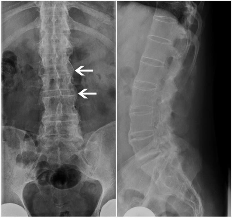
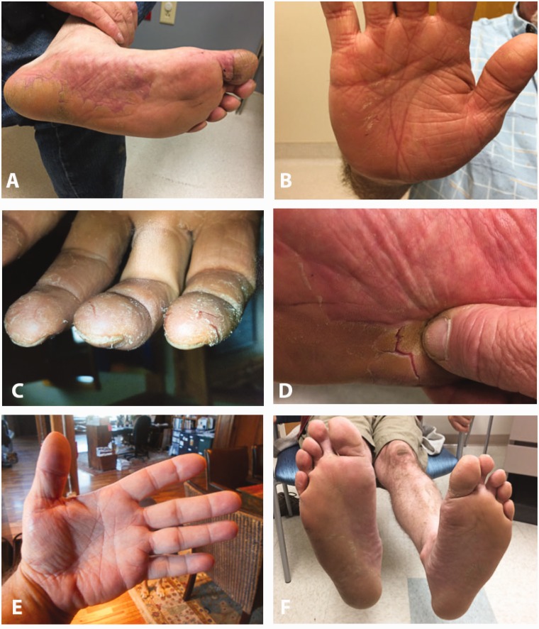
A 56-year-old Caucasian man was referred to the allergy clinic for evaluation of palmoplantar dermatitis. The patient’s rash developed one year prior to presentation. He described erythema, pruritus, and hyperkeratosis of the involved skin with the eventual development of deep fissuring. He had a history of onychomycosis of his toenails but no history of fungal skin rash. He was exposed to solvents, mineral spirits, and gasoline through his occupation in home renovation and wearing neither nitrile nor cotton gloves alleviated his symptoms. Prior evaluations were carried out by primary care and dermatology. He had been treated with topical emollients, topical steroids (including potent agents such as clobetasol) for suspected atopic dermatitis, and topical antifungal agents as well (although KOH prep was negative). All prior treatments failed to resolve his severe palmoplantar rash. His medical history included hypertension, obesity, fatty liver disease, uveitis, bilateral total hip arthroplasty, and a history of childhood allergic rhinitis for which he underwent allergen immunotherapy and was quiescent at the time of evaluation. The physical examination was significant for moderately erythematous, hyperkeratotic, well-defined plaques on the palmoplantar surfaces of the hands and feet without dorsal involvement. Fissuring was seen at the fingertips and the plantar surface of the feet (Figure 1(A) to (D)) without associated pustulosis. There were no obvious nail pitting, oil spots, nor onycholysis, and scalp examination was normal. External examination of the eyes and oral examination were both normal. No appreciable synovitis was documented on peripheral joint examination, but the patient appeared “stiff” with ambulation as well as when stepping down from the examination table. Patch testing result revealed a weak positive reaction to gold sodium thiosulfate and an irritant reaction to thimerosal. Common sensitizers of allergic contact dermatitis in the construction worker were evaluated. The patient did not have reactions to potassium dichromate found in cements, biocides such as isothiazolinones, rubber chemical, and metal allergens (ie, chrome, thiurams, carbamates, mercaptobenzothiazole) accounting for foot dermatitis from work boot materials, and epoxy resin. These results, along with the patient’s history, led to decreased suspicion for either contact or atopic dermatitis. Given the patient’s history of uveitis (which upon review of the chart was recurrent and associated with HLA-B27 positivity), the hyperkeratotic and plaque-like appearance of his lesions, and the concern for possible axial spine disease based on examination, formal radiographs were obtained (Figure 2) and a referral to rheumatology was initiated. His rheumatologic evaluation confirmed decreased range of motion at the spine with an abnormal occiput to wall test of 8 cm, abnormal Schober’s test (10–12 cm increase with flexion and no reversal of lumbar lordosis), and decreased excursion in lateral bending. The constellation of findings was consistent with a diagnosis of axial spondyloarthritis (axial SpA). His skin findings were thought to represent keratoderma blenorrhagicum versus palmoplantar-variant psoriasis (PPP) and were suspected to be a manifestation of his axial SpA. He denied a history of genitourinary or gastrointestinal infection, and urinary testing for gonorrhea and chlamydia was negative. Given failure of two prior


 分享
分享
 求助内容:
求助内容: 应助结果提醒方式:
 扫码关注我们
