Andrei Roman, Patriciu Achimas-Cadariu, Bogdan Fetica, Vlad Gata, Andrada Seicean
{"title":"CT-guided procedures: an initial experience.","authors":"Andrei Roman, Patriciu Achimas-Cadariu, Bogdan Fetica, Vlad Gata, Andrada Seicean","doi":"10.15386/cjmed-1145","DOIUrl":null,"url":null,"abstract":"<p><strong>Background and aims: </strong>Despite their usefulness, CT-guided procedures have a low profile in Romania. The current study has the purpose of describing a first experience in performing these procedures.</p><p><strong>Methods: </strong>Tumors and fluid collections that were inaccessible for biopsy or drainage by ultrasound or endoscopic guidance were included. The procedures were performed using a 64-slice GE Optima CT660 CT scanner. The biopsies were carried out using the coaxial technique with an 18 G semiautomatic needle. The drainages were performed using 10 F pig-tail drains that were inserted using the Seldinger technique. Data regarding the size and location of the target lesion, puncture technique, success and complication rates were recorded.</p><p><strong>Results: </strong>Between May 2017 and April 2018, 30 procedures were performed, of which 26 biopsies and 4 drainage insertions. Of the biopsies 3 were mediastinal, 8 pulmonary, 6 retroperitoneal, 4 pelvic, and 5 of the bone. The drainages were performed for pelvic lymphoceles. The average lesion size was 3.2 cm (0.7-9 cm), with a depth from the skin of 9.1 cm (0.6-15.2 cm). The average procedure duration was 58 minutes (31-93 minutes). A conclusive histopathological diagnosis was set after 92.3% of biopsies. Three procedures resulted in complications, two being minor (hemothorax, soft tissue hematoma) and one severe (tension pneumothorax requiring drainage).</p><p><strong>Conclusions: </strong>CT guidance offers safe access to lesions that cannot be biopsied or drained under ultrasound or endoscopic guidance.</p>","PeriodicalId":91233,"journal":{"name":"Clujul medical (1957)","volume":"91 4","pages":"427-434"},"PeriodicalIF":0.0000,"publicationDate":"2018-10-01","publicationTypes":"Journal Article","fieldsOfStudy":null,"isOpenAccess":false,"openAccessPdf":"https://ftp.ncbi.nlm.nih.gov/pub/pmc/oa_pdf/64/8b/cm-91-427.PMC6296730.pdf","citationCount":"5","resultStr":null,"platform":"Semanticscholar","paperid":null,"PeriodicalName":"Clujul medical (1957)","FirstCategoryId":"1085","ListUrlMain":"https://doi.org/10.15386/cjmed-1145","RegionNum":0,"RegionCategory":null,"ArticlePicture":[],"TitleCN":null,"AbstractTextCN":null,"PMCID":null,"EPubDate":"2018/10/30 0:00:00","PubModel":"Epub","JCR":"","JCRName":"","Score":null,"Total":0}
引用次数: 5
Abstract
Background and aims: Despite their usefulness, CT-guided procedures have a low profile in Romania. The current study has the purpose of describing a first experience in performing these procedures.
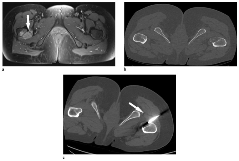
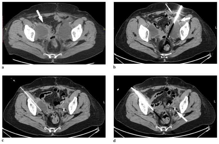
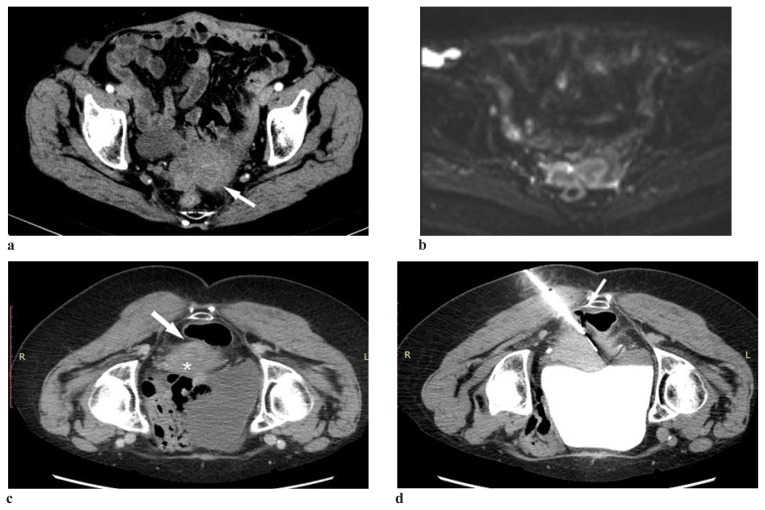
Methods: Tumors and fluid collections that were inaccessible for biopsy or drainage by ultrasound or endoscopic guidance were included. The procedures were performed using a 64-slice GE Optima CT660 CT scanner. The biopsies were carried out using the coaxial technique with an 18 G semiautomatic needle. The drainages were performed using 10 F pig-tail drains that were inserted using the Seldinger technique. Data regarding the size and location of the target lesion, puncture technique, success and complication rates were recorded.
Results: Between May 2017 and April 2018, 30 procedures were performed, of which 26 biopsies and 4 drainage insertions. Of the biopsies 3 were mediastinal, 8 pulmonary, 6 retroperitoneal, 4 pelvic, and 5 of the bone. The drainages were performed for pelvic lymphoceles. The average lesion size was 3.2 cm (0.7-9 cm), with a depth from the skin of 9.1 cm (0.6-15.2 cm). The average procedure duration was 58 minutes (31-93 minutes). A conclusive histopathological diagnosis was set after 92.3% of biopsies. Three procedures resulted in complications, two being minor (hemothorax, soft tissue hematoma) and one severe (tension pneumothorax requiring drainage).
Conclusions: CT guidance offers safe access to lesions that cannot be biopsied or drained under ultrasound or endoscopic guidance.


 分享
分享

 求助内容:
求助内容: 应助结果提醒方式:
 扫码关注我们
