Incremental Role of Fluorine 18-Fluorodeoxyglucose Positron Emission Tomography/Computed Tomography in the Assessment of Computed Tomography-Inconspicuous Pancreatic Lesions.
Fathinul Fikri Ahmad Saad, Anna Misyail Abdul Rashid, Mohamad Syafeeq Faeez Md Noh
{"title":"Incremental Role of Fluorine 18-Fluorodeoxyglucose Positron Emission Tomography/Computed Tomography in the Assessment of Computed Tomography-Inconspicuous Pancreatic Lesions.","authors":"Fathinul Fikri Ahmad Saad, Anna Misyail Abdul Rashid, Mohamad Syafeeq Faeez Md Noh","doi":"10.1089/pancan.2017.0014","DOIUrl":null,"url":null,"abstract":"<p><p><b>Background:</b> Pancreatic malignancies encompass a heterogenous group of disorders, with poor prognosis at diagnosis. Traditionally, conventional computed tomography (CT) has been used for diagnosis, staging, and follow up. However, this technique lacks functional information; and is limited in diagnosis of occult pancreatic disease. Hybrid imaging in the form of positron emission tomography (PET)/CT provides a potential avenue for early detection and subsequent appropriate therapy. <b>Case Presentation:</b> A 60-year-old male, with a history of abdominal aortic aneurysm which was repaired, came with a complaint of 2 months history of back pain, radiating to the front. The pain was relieved on leaning forward, and aggravated by lying on his back. CT angiography of the abdomen was done, which revealed a concealed aortic aneurysm and a significant atrophy of the pancreatic tail. The serum cancer antigen (CA) 19-9 was elevated (50.0 U/mL, reference range 0.0-37.0 U/mL). At this juncture, the PET scan done revealed no discernible abnormalities. Patient was put on close follow-up in view of the rising trend of CA 19-9 levels. Three months following the initial scans, a repeat <sup>18</sup>F-FDG (fluorine 18 fluorodeoxyglucose) PET/CT revealed an FDG-avid lesion at the neck of the pancreas on PET without perceptible changes on the correlated CT. A Whipple's procedure ensued, with histopathological examination findings of pancreatic adenocarcinoma. <b>Conclusion:</b> This article discusses the role of PET/CT in the early diagnosis of inconspicuous pancreatic lesions; which could have averted immediate medical therapy.</p>","PeriodicalId":16655,"journal":{"name":"Journal of Pancreatic Cancer","volume":"3 1","pages":"66-70"},"PeriodicalIF":0.0000,"publicationDate":"2017-09-01","publicationTypes":"Journal Article","fieldsOfStudy":null,"isOpenAccess":false,"openAccessPdf":"https://sci-hub-pdf.com/10.1089/pancan.2017.0014","citationCount":"1","resultStr":null,"platform":"Semanticscholar","paperid":null,"PeriodicalName":"Journal of Pancreatic Cancer","FirstCategoryId":"1085","ListUrlMain":"https://doi.org/10.1089/pancan.2017.0014","RegionNum":0,"RegionCategory":null,"ArticlePicture":[],"TitleCN":null,"AbstractTextCN":null,"PMCID":null,"EPubDate":"2017/1/1 0:00:00","PubModel":"eCollection","JCR":"","JCRName":"","Score":null,"Total":0}
引用次数: 1
Abstract
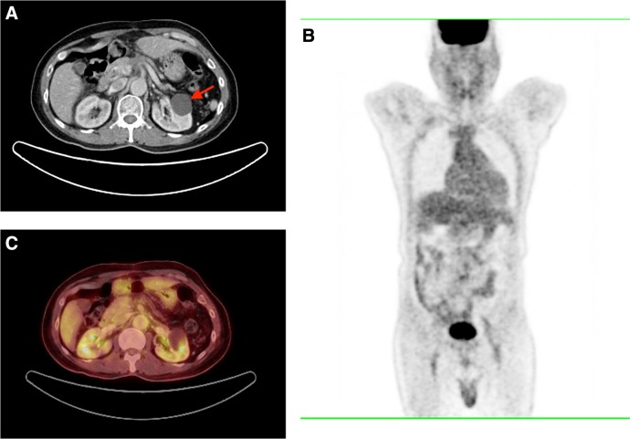
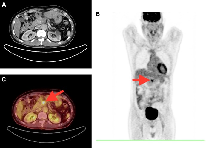
Background: Pancreatic malignancies encompass a heterogenous group of disorders, with poor prognosis at diagnosis. Traditionally, conventional computed tomography (CT) has been used for diagnosis, staging, and follow up. However, this technique lacks functional information; and is limited in diagnosis of occult pancreatic disease. Hybrid imaging in the form of positron emission tomography (PET)/CT provides a potential avenue for early detection and subsequent appropriate therapy. Case Presentation: A 60-year-old male, with a history of abdominal aortic aneurysm which was repaired, came with a complaint of 2 months history of back pain, radiating to the front. The pain was relieved on leaning forward, and aggravated by lying on his back. CT angiography of the abdomen was done, which revealed a concealed aortic aneurysm and a significant atrophy of the pancreatic tail. The serum cancer antigen (CA) 19-9 was elevated (50.0 U/mL, reference range 0.0-37.0 U/mL). At this juncture, the PET scan done revealed no discernible abnormalities. Patient was put on close follow-up in view of the rising trend of CA 19-9 levels. Three months following the initial scans, a repeat 18F-FDG (fluorine 18 fluorodeoxyglucose) PET/CT revealed an FDG-avid lesion at the neck of the pancreas on PET without perceptible changes on the correlated CT. A Whipple's procedure ensued, with histopathological examination findings of pancreatic adenocarcinoma. Conclusion: This article discusses the role of PET/CT in the early diagnosis of inconspicuous pancreatic lesions; which could have averted immediate medical therapy.


 分享
分享

 求助内容:
求助内容: 应助结果提醒方式:
 扫码关注我们
