Axonal changes in experimental prion diseases recapitulate those following constriction of postganglionic branches of the superior cervical ganglion: a comparison 40 years later.
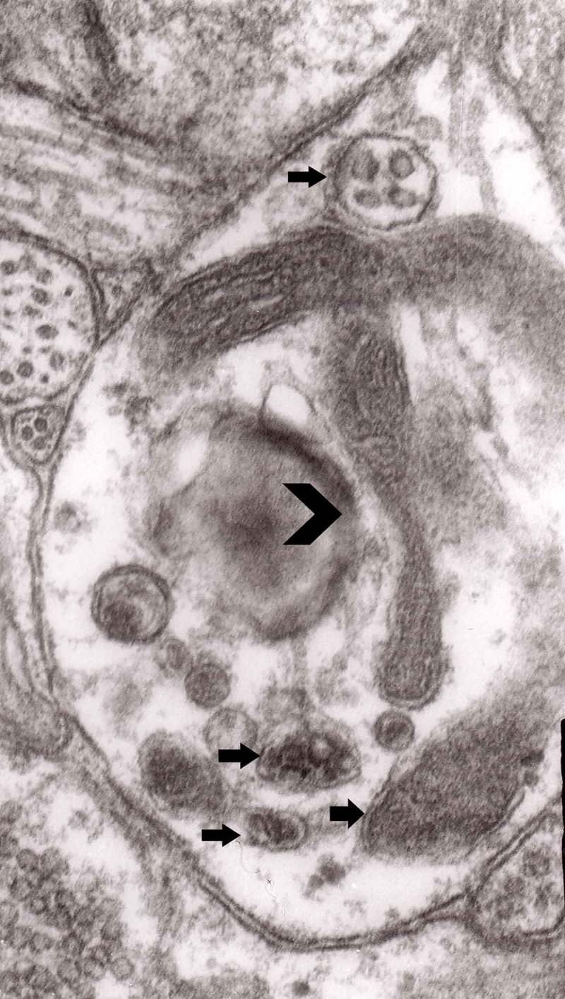
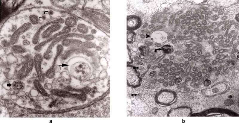
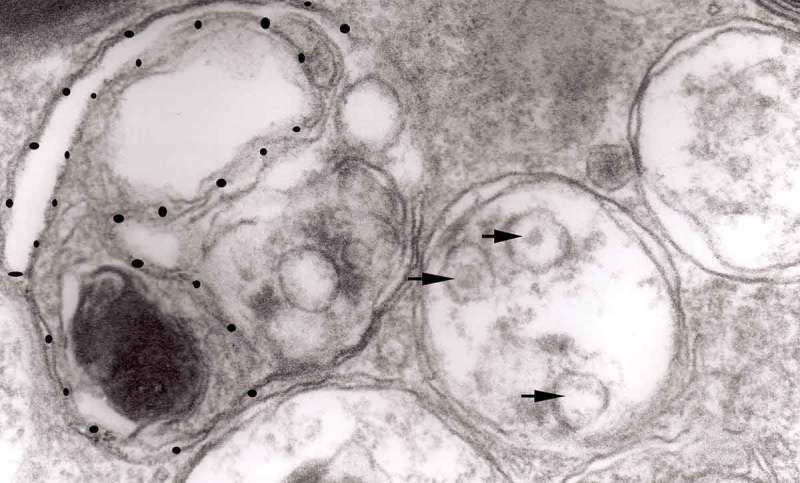
{"title":"Axonal changes in experimental prion diseases recapitulate those following constriction of postganglionic branches of the superior cervical ganglion: a comparison 40 years later.","authors":"Paweł P Liberski","doi":"10.1080/19336896.2019.1595315","DOIUrl":null,"url":null,"abstract":"<p><p>The major neurological feature of prion diseases is a neuronal loss accomplished through either apoptosis or autophagy. In this review, I compared axonal alterations in prion diseases to those described 40 years earlier as a result of nerve ligation. I also demonstrated that autophagic vacuoles and autophagosomes are a major part of dystrophic neurites. Furthermore, I summarized the current status of the autophagy in prion diseases and hypothesize, that spongiform change may originate from the autophagic vacuoles. This conclusion should be supported by other methods, in particular laser confocal microscopy. We observed neuronal autophagic vacuoles in different stages of formation, and our interpretation of the 'maturity' of their formation may or may not equate to actual developmental stages. Initially, a part of the neuronal cytoplasm was sequestrated within double or multiple membranes (phagophores) and often exhibited increased electron-density. The intracytoplasmic membranes formed labyrinth-like structures that suggest a multiplication of those membranes. The autophagic vacuoles then expand and eventually, a vast area of the cytoplasm was transformed into a merging mass of autophagic vacuoles. Margaret R. Matthews published a long treatise in the Philosophical Transactions of the Royal Society of London in which she had described in great detail the ultrastructure of postganglionic branches of the superior cervical ganglion in the rat following ligation of them. The earliest changes observed by Matthews between 6 h to 2 days in the proximal stump were distensions of proximal axons. Analogously, in our models, an increased number of 'regular' (round) and 'irregular' MVB and some autophagic vacuoles were observed collectively, both processes were similar.</p>","PeriodicalId":54585,"journal":{"name":"Prion","volume":"13 1","pages":"83-93"},"PeriodicalIF":1.6000,"publicationDate":"2019-01-01","publicationTypes":"Journal Article","fieldsOfStudy":null,"isOpenAccess":false,"openAccessPdf":"https://sci-hub-pdf.com/10.1080/19336896.2019.1595315","citationCount":"8","resultStr":null,"platform":"Semanticscholar","paperid":null,"PeriodicalName":"Prion","FirstCategoryId":"99","ListUrlMain":"https://doi.org/10.1080/19336896.2019.1595315","RegionNum":3,"RegionCategory":"生物学","ArticlePicture":[],"TitleCN":null,"AbstractTextCN":null,"PMCID":null,"EPubDate":"","PubModel":"","JCR":"Q4","JCRName":"BIOCHEMISTRY & MOLECULAR BIOLOGY","Score":null,"Total":0}
引用次数: 8
Abstract
The major neurological feature of prion diseases is a neuronal loss accomplished through either apoptosis or autophagy. In this review, I compared axonal alterations in prion diseases to those described 40 years earlier as a result of nerve ligation. I also demonstrated that autophagic vacuoles and autophagosomes are a major part of dystrophic neurites. Furthermore, I summarized the current status of the autophagy in prion diseases and hypothesize, that spongiform change may originate from the autophagic vacuoles. This conclusion should be supported by other methods, in particular laser confocal microscopy. We observed neuronal autophagic vacuoles in different stages of formation, and our interpretation of the 'maturity' of their formation may or may not equate to actual developmental stages. Initially, a part of the neuronal cytoplasm was sequestrated within double or multiple membranes (phagophores) and often exhibited increased electron-density. The intracytoplasmic membranes formed labyrinth-like structures that suggest a multiplication of those membranes. The autophagic vacuoles then expand and eventually, a vast area of the cytoplasm was transformed into a merging mass of autophagic vacuoles. Margaret R. Matthews published a long treatise in the Philosophical Transactions of the Royal Society of London in which she had described in great detail the ultrastructure of postganglionic branches of the superior cervical ganglion in the rat following ligation of them. The earliest changes observed by Matthews between 6 h to 2 days in the proximal stump were distensions of proximal axons. Analogously, in our models, an increased number of 'regular' (round) and 'irregular' MVB and some autophagic vacuoles were observed collectively, both processes were similar.
期刊介绍:
Prion is the first international peer-reviewed open access journal to focus exclusively on protein folding and misfolding, protein assembly disorders, protein-based and structural inheritance. The goal is to foster communication and rapid exchange of information through timely publication of important results using traditional as well as electronic formats. The overriding criteria for publication in Prion are originality, scientific merit and general interest.


 分享
分享
 求助内容:
求助内容: 应助结果提醒方式:
 扫码关注我们
