Yeun-Hwa Gu, Takenori Yamashita, Tota Inoue, Jin-Ho Song, Ki-Mun Kang
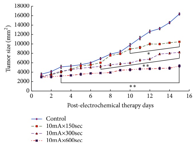
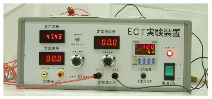
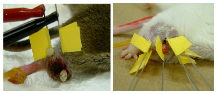
{"title":"Cellular and Molecular Level Mechanisms against Electrochemical Cancer Therapy.","authors":"Yeun-Hwa Gu, Takenori Yamashita, Tota Inoue, Jin-Ho Song, Ki-Mun Kang","doi":"10.1155/2019/3431674","DOIUrl":null,"url":null,"abstract":"<p><p>Electrochemical treatment (ECT) is a promising new way to induce tumor regression by flowing direct current into the cancer tissue. ECT was applied to different kinds of tumors in clinical studies and showed good results. In addition, basic research has almost not been done in the field of evaluation of efficacy, dose-response, and cytotoxicity. Therefore, the objective is to study the cellular mechanism in the antitumor effect of ECT and to contribute data of basic research of ECT. In the cell-level study, tumor cells (Sarcoma-180, Scc-7, Ehrlich Carcinoma) were studied using ICR mice and C3H mice. In the study group, pH values of control, 10mA × 150secs, 10mA × 300secs, and 10mA × 600secs groups were measured five times each. In histological level studies, ECT was performed on tumors inoculated on the upper part of the right foot of C3H mice. In each group, mice were sacrificed by cervical dislocation 6, 12, and 24 hrs after ECT treatment, and tumors were removed. The excised tumor was fixed in tissue with 10% formalin, and HE staining and apoptosis antibody staining were carried out from the obtained tissue section and observation. In the study at the cellular level, statistically significant differences were observed in all ECT groups in Sarcoma in the tumor growth measurement study compared with the control group. Statistically significant differences were also observed in Scc-7 in all ECT groups compared to the control group. In the intratumoral pH measurement study, there was a statistically significant difference between the anode and the cathode in each group compared to the control group. In the examination at the histological level, microscopic observation of a slide stained with apoptosis antibody with a magnification of 400 times showed that 6hrs after ECT it was stronger and then decreased. By performing ECT, a weak current flows in the living body. As a result, changes in tissue pH, generation of gas, etc. occur. In this study, it was also confirmed that the intratumor pH value becomes strongly acidic on the anode side and strongly alkaline on the cathode side. In addition, this study confirmed the occurrence of gas during treatment of ECT. Changes in the pH and the like cause changes in the environment in the cell, denaturation of proteins, apoptosis, and necrosis. In this study, a significant increase in apoptosis was confirmed in each ECT group compared to the control group. Treatment effects by ECT were also observed in tumor growth measurement studies and tumor weight measurement studies. From these research results, ECT is considered to be effective as a tumor treatment method.</p>","PeriodicalId":16788,"journal":{"name":"Journal of Pathogens","volume":"2019 ","pages":"3431674"},"PeriodicalIF":1.1000,"publicationDate":"2019-04-14","publicationTypes":"Journal Article","fieldsOfStudy":null,"isOpenAccess":false,"openAccessPdf":"https://www.ncbi.nlm.nih.gov/pmc/articles/PMC6487119/pdf/","citationCount":"0","resultStr":null,"platform":"Semanticscholar","paperid":null,"PeriodicalName":"Journal of Pathogens","FirstCategoryId":"1085","ListUrlMain":"https://doi.org/10.1155/2019/3431674","RegionNum":0,"RegionCategory":null,"ArticlePicture":[],"TitleCN":null,"AbstractTextCN":null,"PMCID":null,"EPubDate":"2019/1/1 0:00:00","PubModel":"eCollection","JCR":"Q4","JCRName":"MICROBIOLOGY","Score":null,"Total":0}
引用次数: 0
Abstract
Electrochemical treatment (ECT) is a promising new way to induce tumor regression by flowing direct current into the cancer tissue. ECT was applied to different kinds of tumors in clinical studies and showed good results. In addition, basic research has almost not been done in the field of evaluation of efficacy, dose-response, and cytotoxicity. Therefore, the objective is to study the cellular mechanism in the antitumor effect of ECT and to contribute data of basic research of ECT. In the cell-level study, tumor cells (Sarcoma-180, Scc-7, Ehrlich Carcinoma) were studied using ICR mice and C3H mice. In the study group, pH values of control, 10mA × 150secs, 10mA × 300secs, and 10mA × 600secs groups were measured five times each. In histological level studies, ECT was performed on tumors inoculated on the upper part of the right foot of C3H mice. In each group, mice were sacrificed by cervical dislocation 6, 12, and 24 hrs after ECT treatment, and tumors were removed. The excised tumor was fixed in tissue with 10% formalin, and HE staining and apoptosis antibody staining were carried out from the obtained tissue section and observation. In the study at the cellular level, statistically significant differences were observed in all ECT groups in Sarcoma in the tumor growth measurement study compared with the control group. Statistically significant differences were also observed in Scc-7 in all ECT groups compared to the control group. In the intratumoral pH measurement study, there was a statistically significant difference between the anode and the cathode in each group compared to the control group. In the examination at the histological level, microscopic observation of a slide stained with apoptosis antibody with a magnification of 400 times showed that 6hrs after ECT it was stronger and then decreased. By performing ECT, a weak current flows in the living body. As a result, changes in tissue pH, generation of gas, etc. occur. In this study, it was also confirmed that the intratumor pH value becomes strongly acidic on the anode side and strongly alkaline on the cathode side. In addition, this study confirmed the occurrence of gas during treatment of ECT. Changes in the pH and the like cause changes in the environment in the cell, denaturation of proteins, apoptosis, and necrosis. In this study, a significant increase in apoptosis was confirmed in each ECT group compared to the control group. Treatment effects by ECT were also observed in tumor growth measurement studies and tumor weight measurement studies. From these research results, ECT is considered to be effective as a tumor treatment method.


 分享
分享
 求助内容:
求助内容: 应助结果提醒方式:
 扫码关注我们
