Liv Jorunn Høllesli, Martin W Kurz, Gry Inger N Behzadi, Tore Solbakken, Svein Harald Mørkve, Kathinka D Kurz
{"title":"Headache and MRI Changes after Endovascular Treatment of a Cerebral Aneurysm.","authors":"Liv Jorunn Høllesli, Martin W Kurz, Gry Inger N Behzadi, Tore Solbakken, Svein Harald Mørkve, Kathinka D Kurz","doi":"10.1155/2019/6917902","DOIUrl":null,"url":null,"abstract":"<p><strong>Background: </strong>The main complications after endovascular therapy of intracranial aneurysms are aneurysm rupture and thromboembolic events. Yet, the widespread use of magnetic resonance imaging (MRI) in follow-up of these patients also demonstrates other, rarely known complications such as aseptic meningitis and foreign body reaction.</p><p><strong>Case presentation: </strong>A small aneurysm in the right posterior communicating artery was treated with endovascular therapy in a 65 year old woman. Two weeks after successful interventional treatment, the patient developed a headache. On MRI performed five months after intervention, vasogenic edema was seen in the vascular territory of the right internal carotid artery. The edema and the symptoms diminished without specific treatment within a year.</p><p><strong>Interpretation: </strong>The clinical and radiological presentation of this case are suggestive of a foreign body reaction, a treatable condition that radiologists and clinicians should be aware of.</p>","PeriodicalId":30326,"journal":{"name":"Case Reports in Radiology","volume":" ","pages":"6917902"},"PeriodicalIF":0.0000,"publicationDate":"2019-12-20","publicationTypes":"Journal Article","fieldsOfStudy":null,"isOpenAccess":false,"openAccessPdf":"https://sci-hub-pdf.com/10.1155/2019/6917902","citationCount":"1","resultStr":null,"platform":"Semanticscholar","paperid":null,"PeriodicalName":"Case Reports in Radiology","FirstCategoryId":"1085","ListUrlMain":"https://doi.org/10.1155/2019/6917902","RegionNum":0,"RegionCategory":null,"ArticlePicture":[],"TitleCN":null,"AbstractTextCN":null,"PMCID":null,"EPubDate":"2019/1/1 0:00:00","PubModel":"eCollection","JCR":"","JCRName":"","Score":null,"Total":0}
引用次数: 1
Abstract
Background: The main complications after endovascular therapy of intracranial aneurysms are aneurysm rupture and thromboembolic events. Yet, the widespread use of magnetic resonance imaging (MRI) in follow-up of these patients also demonstrates other, rarely known complications such as aseptic meningitis and foreign body reaction.
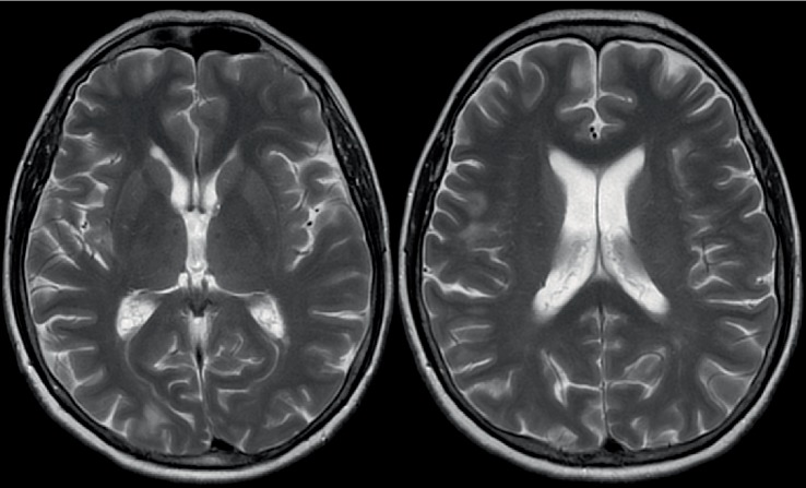
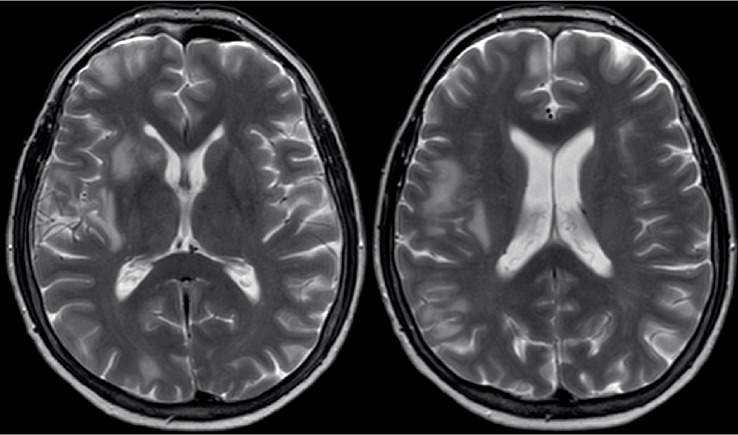
Case presentation: A small aneurysm in the right posterior communicating artery was treated with endovascular therapy in a 65 year old woman. Two weeks after successful interventional treatment, the patient developed a headache. On MRI performed five months after intervention, vasogenic edema was seen in the vascular territory of the right internal carotid artery. The edema and the symptoms diminished without specific treatment within a year.
Interpretation: The clinical and radiological presentation of this case are suggestive of a foreign body reaction, a treatable condition that radiologists and clinicians should be aware of.


 分享
分享

 求助内容:
求助内容: 应助结果提醒方式:
 扫码关注我们
