Eva Gruber-Dujardin, Martina Bleyer, Kerstin Mätz-Rensing
{"title":"Morphological and immunohistochemical characterization of spontaneous endometriosis in rhesus macaques (<i>Macaca mulatta</i>).","authors":"Eva Gruber-Dujardin, Martina Bleyer, Kerstin Mätz-Rensing","doi":"10.5194/pb-4-77-2017","DOIUrl":null,"url":null,"abstract":"<p><p>Several cases of spontaneous endometriosis in middle-aged to old rhesus macaques (<i>Macaca mulatta</i>) from the breeding colony of the German Primate Center were thoroughly characterized with regards to anatomical distribution and macroscopic appearance, histological differentiation and immunohistochemical profile including somatic markers, hormonal receptors, and proliferation indices. More than half of the examined animals (five of nine) were directly related to one breeding male, supporting a strong genetic predisposition. Histologically, four different types of endometriotic lesions, depending on the degree of ectopic endometrial gland and stromal differentiation (well differentiated, purely stromal, mixed differentiation, poorly differentiated), could be constantly identified within all animals. Immunohistochemistry (IHC) of cytokeratin (CK), vimentin, smooth muscle actin (SMA), desmin, estrogen (ER), and progesterone (PR) receptors as well as of the nuclear proteins Ki67 and p53 revealed varying staining patterns in the four different types of endometriosis differentiation and compared to normal endometrium. Purely stromal, mixed, or poorly differentiated lesions, especially, showed additional cytokeratin-positive stromal cells, whereas epithelial cells of endometriosis with mixed or poor differentiation increasingly expressed mesenchymal markers (vimentin, SMA). Hormonal receptor and Ki67 expression in well-differentiated endometriotic lesions mostly reflected that of normal endometrial tissue according to the cyclic phase of the animal, while the expression gradually diminished with decreasing grade of differentiation. However, increased nuclear accumulations of p53 antigen could only be continuously detected in epithelial cells of mixed or poorly differentiated endometriosis. Altogether, these findings support the pathogenetic theory of coelomic metaplasia, since the expression profiles of somatic markers in less differentiated forms closely resembled that of mesothelial cells. Thus, the four different histological types of endometriosis might display subsequent grades of differentiation in the course of time, with poorly differentiated types representing newly formed, immature lesions and well-differentiated types being older, fully differentiated forms, rather than being the outcome of dedifferentiation processes.</p>","PeriodicalId":37245,"journal":{"name":"Primate Biology","volume":"4 1","pages":"77-91"},"PeriodicalIF":0.0000,"publicationDate":"2017-04-13","publicationTypes":"Journal Article","fieldsOfStudy":null,"isOpenAccess":false,"openAccessPdf":"https://www.ncbi.nlm.nih.gov/pmc/articles/PMC7041517/pdf/","citationCount":"0","resultStr":null,"platform":"Semanticscholar","paperid":null,"PeriodicalName":"Primate Biology","FirstCategoryId":"1085","ListUrlMain":"https://doi.org/10.5194/pb-4-77-2017","RegionNum":0,"RegionCategory":null,"ArticlePicture":[],"TitleCN":null,"AbstractTextCN":null,"PMCID":null,"EPubDate":"2017/1/1 0:00:00","PubModel":"eCollection","JCR":"Q2","JCRName":"Agricultural and Biological Sciences","Score":null,"Total":0}
引用次数: 0
Abstract
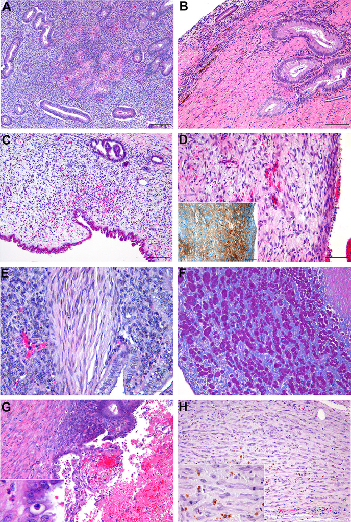
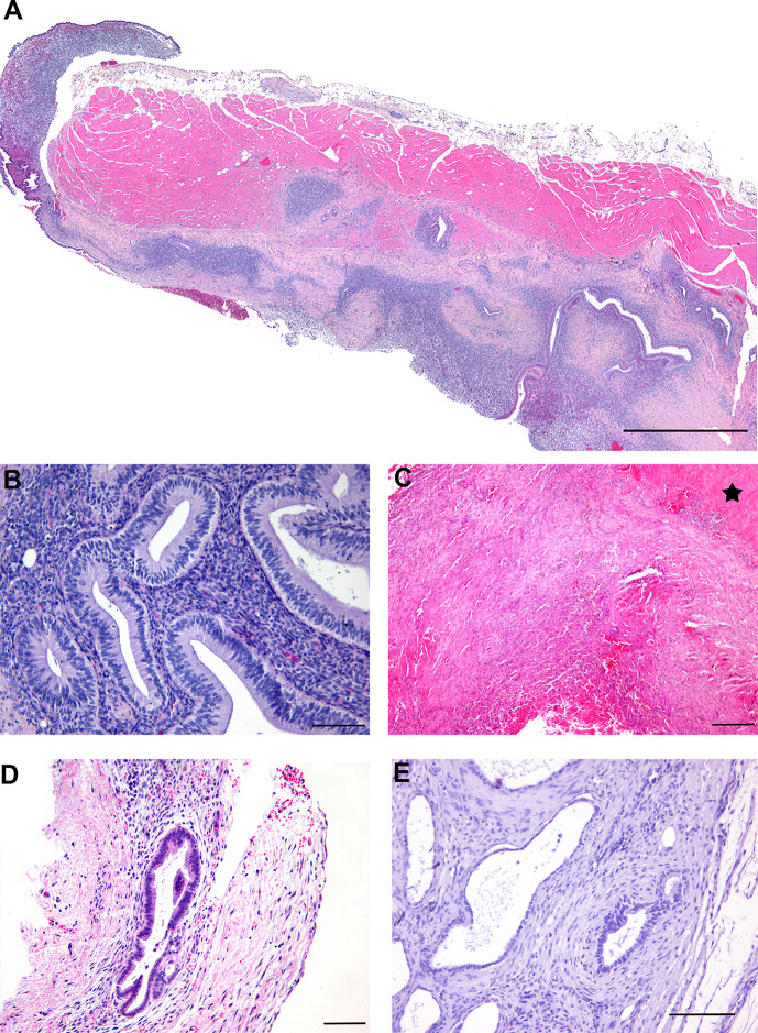
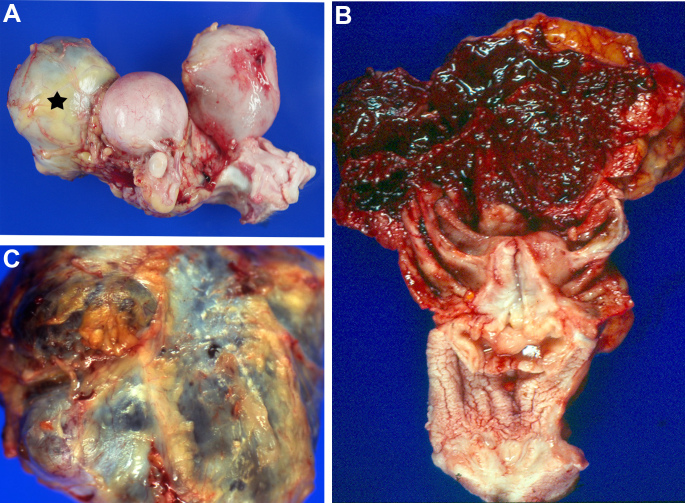
Several cases of spontaneous endometriosis in middle-aged to old rhesus macaques (Macaca mulatta) from the breeding colony of the German Primate Center were thoroughly characterized with regards to anatomical distribution and macroscopic appearance, histological differentiation and immunohistochemical profile including somatic markers, hormonal receptors, and proliferation indices. More than half of the examined animals (five of nine) were directly related to one breeding male, supporting a strong genetic predisposition. Histologically, four different types of endometriotic lesions, depending on the degree of ectopic endometrial gland and stromal differentiation (well differentiated, purely stromal, mixed differentiation, poorly differentiated), could be constantly identified within all animals. Immunohistochemistry (IHC) of cytokeratin (CK), vimentin, smooth muscle actin (SMA), desmin, estrogen (ER), and progesterone (PR) receptors as well as of the nuclear proteins Ki67 and p53 revealed varying staining patterns in the four different types of endometriosis differentiation and compared to normal endometrium. Purely stromal, mixed, or poorly differentiated lesions, especially, showed additional cytokeratin-positive stromal cells, whereas epithelial cells of endometriosis with mixed or poor differentiation increasingly expressed mesenchymal markers (vimentin, SMA). Hormonal receptor and Ki67 expression in well-differentiated endometriotic lesions mostly reflected that of normal endometrial tissue according to the cyclic phase of the animal, while the expression gradually diminished with decreasing grade of differentiation. However, increased nuclear accumulations of p53 antigen could only be continuously detected in epithelial cells of mixed or poorly differentiated endometriosis. Altogether, these findings support the pathogenetic theory of coelomic metaplasia, since the expression profiles of somatic markers in less differentiated forms closely resembled that of mesothelial cells. Thus, the four different histological types of endometriosis might display subsequent grades of differentiation in the course of time, with poorly differentiated types representing newly formed, immature lesions and well-differentiated types being older, fully differentiated forms, rather than being the outcome of dedifferentiation processes.


 分享
分享
 求助内容:
求助内容: 应助结果提醒方式:
 扫码关注我们
