Gayane Manukyan, Anush Martirosyan, Ludek Slavik, Sona Margaryan, Jana Ulehlova, Zuzana Mikulkova, Antonin Hlusi, Tomas Papajik, Eva Kriegova
{"title":"Anti-domain 1 β2 glycoprotein antibodies increase expression of tissue factor on monocytes and activate NK Cells and CD8+ cells in vitro.","authors":"Gayane Manukyan, Anush Martirosyan, Ludek Slavik, Sona Margaryan, Jana Ulehlova, Zuzana Mikulkova, Antonin Hlusi, Tomas Papajik, Eva Kriegova","doi":"10.1186/s13317-020-00128-y","DOIUrl":null,"url":null,"abstract":"<p><strong>Background: </strong>β2-Glycoprotein I (β2GPI) represents the major antigenic target for antiphospholipid antibodies (aPL), with domain 1 (D1) being identified as a risk factor for thrombosis and pregnancy complications in APS. We aimed to analyse the ability of aPL, and particularly anti-D1 β2GPI, to stimulate prothrombotic and proinflammatory activity of immune cells in vitro.</p><p><strong>Methods: </strong>Peripheral blood mononuclear cells (PBMCs) from 11 healthy individuals were incubated with: (1) \"anti-D1(+)\"-pooled plasma derived from patients suspected of having APS contained anticardiolipin antibodies (aCL), lupus anticoagulant (LA), anti-β2GPI and anti-D1 β2GPI; (2) \"anti-D1(-)\"-pooled plasma from patients suspected of having APS contained aCL, LA, anti-β2GPI, and negative for anti-D1 β2GPI; (3) \"seronegative\"-negative for aPL.</p><p><strong>Results: </strong>The presence of anti-D1(+) and anti-D1(-) plasma resulted in increased HLA-DR and CD11b on monocytes. While only anti-D1(+) plasma markedly increased the percentage and median fluorescence intensity (MFI) of CD142 (tissue factor, TF) on monocytes in comparison with those cultured with anti-D1(-) and seronegative plasma. Anti-D1(+) plasma resulted in increased percentage and MFI of activation marker CD69 on NK and T cytotoxic cells. Expression of IgG receptor FcγRIII(CD16) on monocytes and NK cells was down-regulated by the anti-D1(+) plasma.</p><p><strong>Conclusions: </strong>Taking together, our study shows the ability of patient-derived aPL to induce immune cell activation and TF expression on monocytes. For the first time, we demonstrated the influence of anti-D1 β2GPI on the activation status of monocytes, NK and cytotoxic T cells. Our findings further support a crucial role of D1 epitope in the promotion of thrombosis and obstetrical complications in APS.</p>","PeriodicalId":8655,"journal":{"name":"Auto-Immunity Highlights","volume":" ","pages":"5"},"PeriodicalIF":0.0000,"publicationDate":"2020-03-02","publicationTypes":"Journal Article","fieldsOfStudy":null,"isOpenAccess":false,"openAccessPdf":"https://sci-hub-pdf.com/10.1186/s13317-020-00128-y","citationCount":"6","resultStr":null,"platform":"Semanticscholar","paperid":null,"PeriodicalName":"Auto-Immunity Highlights","FirstCategoryId":"1085","ListUrlMain":"https://doi.org/10.1186/s13317-020-00128-y","RegionNum":0,"RegionCategory":null,"ArticlePicture":[],"TitleCN":null,"AbstractTextCN":null,"PMCID":null,"EPubDate":"","PubModel":"","JCR":"Q1","JCRName":"Medicine","Score":null,"Total":0}
引用次数: 6
Abstract
Background: β2-Glycoprotein I (β2GPI) represents the major antigenic target for antiphospholipid antibodies (aPL), with domain 1 (D1) being identified as a risk factor for thrombosis and pregnancy complications in APS. We aimed to analyse the ability of aPL, and particularly anti-D1 β2GPI, to stimulate prothrombotic and proinflammatory activity of immune cells in vitro.
Methods: Peripheral blood mononuclear cells (PBMCs) from 11 healthy individuals were incubated with: (1) "anti-D1(+)"-pooled plasma derived from patients suspected of having APS contained anticardiolipin antibodies (aCL), lupus anticoagulant (LA), anti-β2GPI and anti-D1 β2GPI; (2) "anti-D1(-)"-pooled plasma from patients suspected of having APS contained aCL, LA, anti-β2GPI, and negative for anti-D1 β2GPI; (3) "seronegative"-negative for aPL.
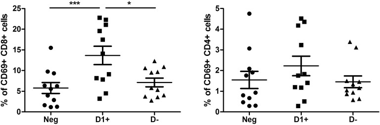
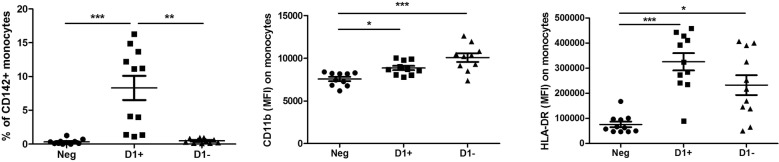
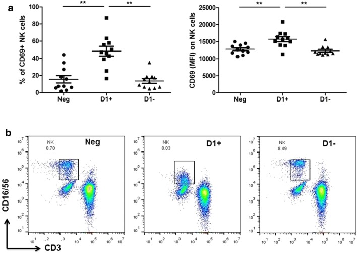
Results: The presence of anti-D1(+) and anti-D1(-) plasma resulted in increased HLA-DR and CD11b on monocytes. While only anti-D1(+) plasma markedly increased the percentage and median fluorescence intensity (MFI) of CD142 (tissue factor, TF) on monocytes in comparison with those cultured with anti-D1(-) and seronegative plasma. Anti-D1(+) plasma resulted in increased percentage and MFI of activation marker CD69 on NK and T cytotoxic cells. Expression of IgG receptor FcγRIII(CD16) on monocytes and NK cells was down-regulated by the anti-D1(+) plasma.
Conclusions: Taking together, our study shows the ability of patient-derived aPL to induce immune cell activation and TF expression on monocytes. For the first time, we demonstrated the influence of anti-D1 β2GPI on the activation status of monocytes, NK and cytotoxic T cells. Our findings further support a crucial role of D1 epitope in the promotion of thrombosis and obstetrical complications in APS.


 分享
分享
 求助内容:
求助内容: 应助结果提醒方式:
 扫码关注我们
