{"title":"Mature Ovarian Teratoma: Atypical Imaging.","authors":"S Wakrim, M El Jdid","doi":"10.1155/2020/1352961","DOIUrl":null,"url":null,"abstract":"<p><p>The incidence of a mature ovarian teratoma ranged from 20% to 30% of pediatric ovarian tumors (Sabaa et al., 2009), which is composed of well-differentiated tissues that derive from all three germ cell layers (ectoderm, mesoderm, and endoderm); it is one of the most common benign ovarian neoplasms. In this case report, we discuss a 9-year-old female patient who presented with abdominal pain and distended abdomen, for which she had an abdominal ultrasound and magnetic resonance imaging. The histopathological exam, after a laparotomy, showed a mature ovarian teratoma.</p>","PeriodicalId":30326,"journal":{"name":"Case Reports in Radiology","volume":"2020 ","pages":"1352961"},"PeriodicalIF":0.0000,"publicationDate":"2020-02-18","publicationTypes":"Journal Article","fieldsOfStudy":null,"isOpenAccess":false,"openAccessPdf":"https://sci-hub-pdf.com/10.1155/2020/1352961","citationCount":"0","resultStr":null,"platform":"Semanticscholar","paperid":null,"PeriodicalName":"Case Reports in Radiology","FirstCategoryId":"1085","ListUrlMain":"https://doi.org/10.1155/2020/1352961","RegionNum":0,"RegionCategory":null,"ArticlePicture":[],"TitleCN":null,"AbstractTextCN":null,"PMCID":null,"EPubDate":"2020/1/1 0:00:00","PubModel":"eCollection","JCR":"","JCRName":"","Score":null,"Total":0}
引用次数: 0
Abstract
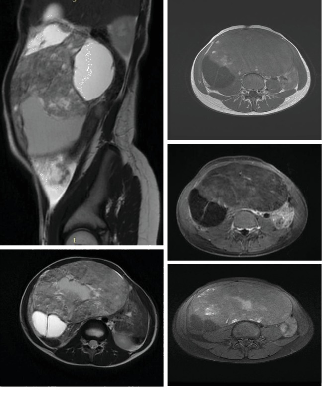
The incidence of a mature ovarian teratoma ranged from 20% to 30% of pediatric ovarian tumors (Sabaa et al., 2009), which is composed of well-differentiated tissues that derive from all three germ cell layers (ectoderm, mesoderm, and endoderm); it is one of the most common benign ovarian neoplasms. In this case report, we discuss a 9-year-old female patient who presented with abdominal pain and distended abdomen, for which she had an abdominal ultrasound and magnetic resonance imaging. The histopathological exam, after a laparotomy, showed a mature ovarian teratoma.


 分享
分享

 求助内容:
求助内容: 应助结果提醒方式:
 扫码关注我们
