Esteban Jaramillo-Jiménez, Manu Gupta, George Snipes, Brennen S Cheek, Christopher B Michael, Ana M Navarro-Montoya, Tatiana Gómez-Escobar, Juliana Jiménez-Villegas, Iader Rodríguez-Márquez, Isaac Melguizo-Gavilanes
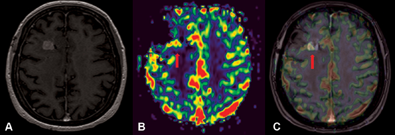
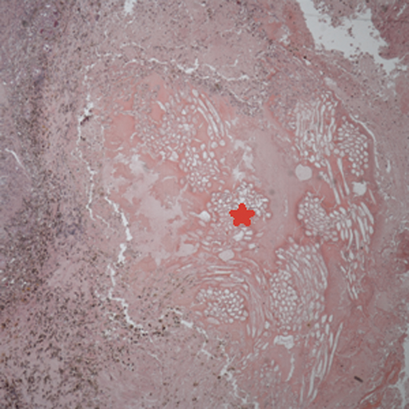
{"title":"Textiloma Mimicking a Recurrent High-Grade Astrocytoma: A Case Report.","authors":"Esteban Jaramillo-Jiménez, Manu Gupta, George Snipes, Brennen S Cheek, Christopher B Michael, Ana M Navarro-Montoya, Tatiana Gómez-Escobar, Juliana Jiménez-Villegas, Iader Rodríguez-Márquez, Isaac Melguizo-Gavilanes","doi":"10.1055/s-0039-3400231","DOIUrl":null,"url":null,"abstract":"<p><p><b>Introduction</b> Textiloma (Txm) is a nonmedical term that has been given to foreign body-related inflammatory pseudotumor arising from retained nonabsorbable cotton matrix that is either inadvertently or deliberately left behind during surgery, which may trigger an inflammatory reaction. This report describes a case of Txm mimicking a recurrent high-grade astrocytoma. <b>Case Report</b> We, here, present the case of a 69-year-old female with a 6-month history of progressive left-sided weakness. Neuroimaging studies revealed a large nonenhancing mass in the right frontoparietal lobe. Pathology reported a World Health Organization tumor classification grade II, diffuse astrocytoma. After surgical intervention, external beam radiation was given to the remaining areas of residual tumor. Routine magnetic resonance imaging (MRI) revealed a nodular area of contrast enhancement in the dorsal and inferior margin of the biopsy tract, growing between interval scans, and perfusion-weighted imaging parameters were elevated being clinically asymptomatic. She underwent a complete resection of this area of interest and pathology returned as a Txm with Surgicel fibers. <b>Conclusion</b> After treatment of a neoplasm, if unexpected clinical or imaging evidence of recurrence is present, a foreign body reaction to hemostatic material used during the initial surgery should be included in the differential diagnosis.</p>","PeriodicalId":44256,"journal":{"name":"Journal of Neurological Surgery Reports","volume":"81 1","pages":"e7-e9"},"PeriodicalIF":0.7000,"publicationDate":"2020-01-01","publicationTypes":"Journal Article","fieldsOfStudy":null,"isOpenAccess":false,"openAccessPdf":"https://sci-hub-pdf.com/10.1055/s-0039-3400231","citationCount":"3","resultStr":null,"platform":"Semanticscholar","paperid":null,"PeriodicalName":"Journal of Neurological Surgery Reports","FirstCategoryId":"1085","ListUrlMain":"https://doi.org/10.1055/s-0039-3400231","RegionNum":0,"RegionCategory":null,"ArticlePicture":[],"TitleCN":null,"AbstractTextCN":null,"PMCID":null,"EPubDate":"2020/3/22 0:00:00","PubModel":"Epub","JCR":"Q4","JCRName":"CLINICAL NEUROLOGY","Score":null,"Total":0}
引用次数: 3
Abstract
Introduction Textiloma (Txm) is a nonmedical term that has been given to foreign body-related inflammatory pseudotumor arising from retained nonabsorbable cotton matrix that is either inadvertently or deliberately left behind during surgery, which may trigger an inflammatory reaction. This report describes a case of Txm mimicking a recurrent high-grade astrocytoma. Case Report We, here, present the case of a 69-year-old female with a 6-month history of progressive left-sided weakness. Neuroimaging studies revealed a large nonenhancing mass in the right frontoparietal lobe. Pathology reported a World Health Organization tumor classification grade II, diffuse astrocytoma. After surgical intervention, external beam radiation was given to the remaining areas of residual tumor. Routine magnetic resonance imaging (MRI) revealed a nodular area of contrast enhancement in the dorsal and inferior margin of the biopsy tract, growing between interval scans, and perfusion-weighted imaging parameters were elevated being clinically asymptomatic. She underwent a complete resection of this area of interest and pathology returned as a Txm with Surgicel fibers. Conclusion After treatment of a neoplasm, if unexpected clinical or imaging evidence of recurrence is present, a foreign body reaction to hemostatic material used during the initial surgery should be included in the differential diagnosis.


 分享
分享
 求助内容:
求助内容: 应助结果提醒方式:
 扫码关注我们
