Proptosis, Orbital Pain, and Long-Standing Monocular Vision Loss Resolved by Surgical Resection of Intraosseous Spheno-Orbital Meningioma: A Case Report and Literature Review.
Jonathan M Parish, Michael Shields, Mackenzie Jones, Scott D Wait, Vinay R Deshmukh
{"title":"Proptosis, Orbital Pain, and Long-Standing Monocular Vision Loss Resolved by Surgical Resection of Intraosseous Spheno-Orbital Meningioma: A Case Report and Literature Review.","authors":"Jonathan M Parish, Michael Shields, Mackenzie Jones, Scott D Wait, Vinay R Deshmukh","doi":"10.1055/s-0040-1708845","DOIUrl":null,"url":null,"abstract":"<p><p><b>Background and Importance</b> We present a case of a patient with a residual intraosseous sphenoid wing meningioma presenting with proptosis, orbital pain, and monocular vision loss for 8 months who underwent decompression of the optic canal, orbital contents, and orbital reconstruction resulting in significant improvement in her vision loss with full resolution of proptosis and orbital pain. <b>Clinical Presentation</b> A 43-year-old female presented with a 1 year history of headache, peri-orbital pain, proptosis, and severe vision loss. She had previously undergone subtotal resection of a large Simpson Grade 1 spheno-orbital meningioma 3 years prior at an outside institution. Workup at our institution revealed hyperostosis of the left greater wing of the sphenoid bone and narrowing of the optic canal along with bony enhancement concerning for residual tumor. The patient was given the recommendation from outside institutions for radiation, presumably due to the chronicity of her visual loss. Our institution recommended resection of the residual osseous tumor with orbital reconstruction. Less than 2 weeks after surgery, the patient noted significant improvement in orbital pain and vision. At 3 months, she had regained full and symmetric orbital appearance with no orbital pain. Her visual acuity improved to 20/30 with full visual fields. <b>Conclusion</b> Surgical decompression of the optic canal and orbital contents for tumor related sphenoid wing hyperostosis should be strongly considered, despite an extended duration of visual change and loss. This case report shows that vision can be significantly restored even after symptoms have been present for greater than 6 months.</p>","PeriodicalId":44256,"journal":{"name":"Journal of Neurological Surgery Reports","volume":"81 1","pages":"e28-e32"},"PeriodicalIF":0.7000,"publicationDate":"2020-01-01","publicationTypes":"Journal Article","fieldsOfStudy":null,"isOpenAccess":false,"openAccessPdf":"https://sci-hub-pdf.com/10.1055/s-0040-1708845","citationCount":"4","resultStr":null,"platform":"Semanticscholar","paperid":null,"PeriodicalName":"Journal of Neurological Surgery Reports","FirstCategoryId":"1085","ListUrlMain":"https://doi.org/10.1055/s-0040-1708845","RegionNum":0,"RegionCategory":null,"ArticlePicture":[],"TitleCN":null,"AbstractTextCN":null,"PMCID":null,"EPubDate":"2020/3/31 0:00:00","PubModel":"Epub","JCR":"Q4","JCRName":"CLINICAL NEUROLOGY","Score":null,"Total":0}
引用次数: 4
Abstract
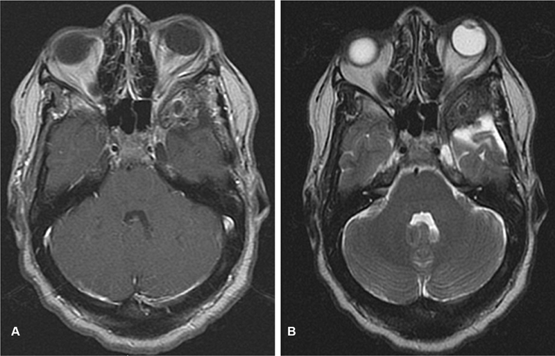
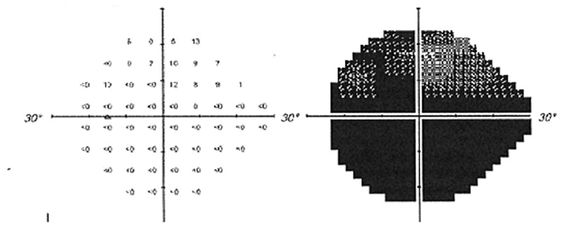
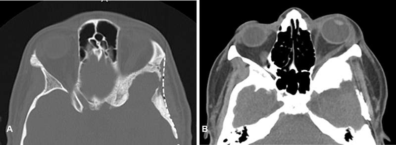
Background and Importance We present a case of a patient with a residual intraosseous sphenoid wing meningioma presenting with proptosis, orbital pain, and monocular vision loss for 8 months who underwent decompression of the optic canal, orbital contents, and orbital reconstruction resulting in significant improvement in her vision loss with full resolution of proptosis and orbital pain. Clinical Presentation A 43-year-old female presented with a 1 year history of headache, peri-orbital pain, proptosis, and severe vision loss. She had previously undergone subtotal resection of a large Simpson Grade 1 spheno-orbital meningioma 3 years prior at an outside institution. Workup at our institution revealed hyperostosis of the left greater wing of the sphenoid bone and narrowing of the optic canal along with bony enhancement concerning for residual tumor. The patient was given the recommendation from outside institutions for radiation, presumably due to the chronicity of her visual loss. Our institution recommended resection of the residual osseous tumor with orbital reconstruction. Less than 2 weeks after surgery, the patient noted significant improvement in orbital pain and vision. At 3 months, she had regained full and symmetric orbital appearance with no orbital pain. Her visual acuity improved to 20/30 with full visual fields. Conclusion Surgical decompression of the optic canal and orbital contents for tumor related sphenoid wing hyperostosis should be strongly considered, despite an extended duration of visual change and loss. This case report shows that vision can be significantly restored even after symptoms have been present for greater than 6 months.


 分享
分享
 求助内容:
求助内容: 应助结果提醒方式:
 扫码关注我们
