{"title":"Assessment of prevalence and position of mandibular incisive canal: A cone beam computed tomography study.","authors":"Reema Talat Ayesha, Balaji Pachipulusu, Poornima Govindaraju","doi":"10.4103/tcmj.tcmj_76_19","DOIUrl":null,"url":null,"abstract":"<p><strong>Objectives: </strong>To avoid anatomical and functional damage to mandibular interforaminal region during surgeries, it is necessary to detect the existence of mandibular incisive canal (MIC) and its proximity to adjacent structures. This study was aimed to evaluate the prevalence of MIC and its proximity to adjacent structures among Indian population.</p><p><strong>Materials and methods: </strong>The images of 80 subjects with the age range of 20-60 years who had undergone cone beam computed tomography examination of the mandible were retrieved from the archival records. There was equal distribution of males and females.</p><p><strong>Results: </strong>The prevalence of MIC in the current study sample was found to be 43.89% with a slightly higher prevalence on left side as compared to right side, and higher prevalence among females as compared to males. Among different age groups, there was an increased incidence in the age group of >50 years. The distance of MIC from labial and lingual cortical plates and lower border of mandible were 4.338 ± 1.478 mm, 4.34 ± 1.53 mm and 9.417 ± 1.832 mm respectively.</p><p><strong>Conclusions: </strong>To conclude, the prevalence of MIC among Indian population was lower as compared to the prevalence among other populations. There were variations in prevalence in terms of age, gender and laterality, which could be used as a reference for further studies conducted on larger sample size. Mapping the incisive nerve canal will enable oral radiologists, to plan safely and negotiate the interforaminal region.</p>","PeriodicalId":72593,"journal":{"name":"Ci ji yi xue za zhi = Tzu-chi medical journal","volume":"32 2","pages":"205-210"},"PeriodicalIF":0.0000,"publicationDate":"2019-09-19","publicationTypes":"Journal Article","fieldsOfStudy":null,"isOpenAccess":false,"openAccessPdf":"https://ftp.ncbi.nlm.nih.gov/pub/pmc/oa_pdf/5a/33/TCMJ-32-205.PMC7137369.pdf","citationCount":"0","resultStr":null,"platform":"Semanticscholar","paperid":null,"PeriodicalName":"Ci ji yi xue za zhi = Tzu-chi medical journal","FirstCategoryId":"1085","ListUrlMain":"https://doi.org/10.4103/tcmj.tcmj_76_19","RegionNum":0,"RegionCategory":null,"ArticlePicture":[],"TitleCN":null,"AbstractTextCN":null,"PMCID":null,"EPubDate":"2020/4/1 0:00:00","PubModel":"eCollection","JCR":"","JCRName":"","Score":null,"Total":0}
引用次数: 0
Abstract
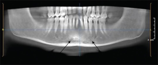
Objectives: To avoid anatomical and functional damage to mandibular interforaminal region during surgeries, it is necessary to detect the existence of mandibular incisive canal (MIC) and its proximity to adjacent structures. This study was aimed to evaluate the prevalence of MIC and its proximity to adjacent structures among Indian population.
Materials and methods: The images of 80 subjects with the age range of 20-60 years who had undergone cone beam computed tomography examination of the mandible were retrieved from the archival records. There was equal distribution of males and females.
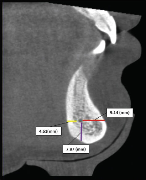
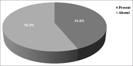
Results: The prevalence of MIC in the current study sample was found to be 43.89% with a slightly higher prevalence on left side as compared to right side, and higher prevalence among females as compared to males. Among different age groups, there was an increased incidence in the age group of >50 years. The distance of MIC from labial and lingual cortical plates and lower border of mandible were 4.338 ± 1.478 mm, 4.34 ± 1.53 mm and 9.417 ± 1.832 mm respectively.
Conclusions: To conclude, the prevalence of MIC among Indian population was lower as compared to the prevalence among other populations. There were variations in prevalence in terms of age, gender and laterality, which could be used as a reference for further studies conducted on larger sample size. Mapping the incisive nerve canal will enable oral radiologists, to plan safely and negotiate the interforaminal region.


 分享
分享

 求助内容:
求助内容: 应助结果提醒方式:
 扫码关注我们
