{"title":"Nanoscale Imaging of Synaptic Connections with Expansion Microscopy.","authors":"Brendan Gallagher, Yongxin Zhao","doi":"10.15190/d.2019.14","DOIUrl":null,"url":null,"abstract":"<p><p>Biologists have long looked for ways to circumvent the physical diffraction limit of light and have developed many strategies to accomplish this. While many techniques employed to image sub-diffraction-limit structures rely on sophisticated equipment and computational methods, expansion microscopy (ExM) is unique in that it provides increase in resolution by physically expanding the sample embedded in a water-swellable hydrogel. ExM has rapidly grown in prevalence, owing to its ease of use and economic nature - all necessary reagents are commercially available, and samples may be imaged in large volume on conventional fluorescence microscopes. Here, we demonstrate the power of expansion microscopy on imaging synaptic connections onto a dopaminergic neuron, in the mouse substantia nigra pars compacta, with nanoscale resolution.</p>","PeriodicalId":72829,"journal":{"name":"Discoveries (Craiova, Romania)","volume":"7 3","pages":"e101"},"PeriodicalIF":0.0000,"publicationDate":"2019-09-30","publicationTypes":"Journal Article","fieldsOfStudy":null,"isOpenAccess":false,"openAccessPdf":"https://www.ncbi.nlm.nih.gov/pmc/articles/PMC7093071/pdf/","citationCount":"3","resultStr":null,"platform":"Semanticscholar","paperid":null,"PeriodicalName":"Discoveries (Craiova, Romania)","FirstCategoryId":"1085","ListUrlMain":"https://doi.org/10.15190/d.2019.14","RegionNum":0,"RegionCategory":null,"ArticlePicture":[],"TitleCN":null,"AbstractTextCN":null,"PMCID":null,"EPubDate":"","PubModel":"","JCR":"","JCRName":"","Score":null,"Total":0}
引用次数: 3
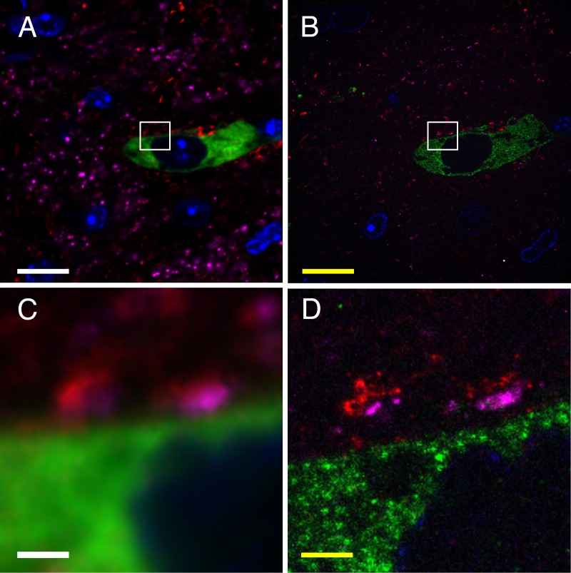
Abstract
Biologists have long looked for ways to circumvent the physical diffraction limit of light and have developed many strategies to accomplish this. While many techniques employed to image sub-diffraction-limit structures rely on sophisticated equipment and computational methods, expansion microscopy (ExM) is unique in that it provides increase in resolution by physically expanding the sample embedded in a water-swellable hydrogel. ExM has rapidly grown in prevalence, owing to its ease of use and economic nature - all necessary reagents are commercially available, and samples may be imaged in large volume on conventional fluorescence microscopes. Here, we demonstrate the power of expansion microscopy on imaging synaptic connections onto a dopaminergic neuron, in the mouse substantia nigra pars compacta, with nanoscale resolution.


 分享
分享

 求助内容:
求助内容: 应助结果提醒方式:
 扫码关注我们
