Unilateral maxillary central incisor root resorption after orthodontic treatment for Angle Class II, division 1 malocclusion with significant maxillary midline deviation: A possible correlation with root proximity to the incisive canal.
{"title":"Unilateral maxillary central incisor root resorption after orthodontic treatment for Angle Class II, division 1 malocclusion with significant maxillary midline deviation: A possible correlation with root proximity to the incisive canal.","authors":"Toshihiro Imamura, Shunsuke Uesugi, Takashi Ono","doi":"10.4041/kjod.2020.50.3.216","DOIUrl":null,"url":null,"abstract":"<p><p>Root resorption can be caused by several factors, including contact with the cortical bone. Here we report a case involving a 21-year-old female with Angle Class II, division 1 malocclusion who exhibited significant root resorption in the maxillary right central incisor after orthodontic treatment. The patient presented with significant left-sided deviation of the maxillary incisors due to lingual dislocation of the left lateral incisor and a Class II molar relationship. Cephalometric analysis demonstrated a Class I skeletal relationship (A pointnasion-B point, 2.5°) and proclined maxillary anterior teeth (upper incisor to sella-nasion plane angle, 113.4°). The primary treatment objectives were the achievement of stable occlusion with midline agreement between the maxillary and mandibular dentitions and appropriate maxillary anterior tooth axes and molar relationship. A panoramic radiograph obtained after active treatment showed significant root resorption in the maxillary right central incisor; therefore, we performed cone-beam computed tomography, which confirmed root resorption along the cortical bone around the incisive canal. The findings from this case, where different degrees of root resorption were observed despite comparable degrees of orthodontic movement in the bilateral maxillary central incisors, suggest that the incisive canal could be an inducing factor for root resorption. However, further investigation is necessary to confirm this assumption.</p>","PeriodicalId":49934,"journal":{"name":"Korean Journal of Orthodontics","volume":"50 3","pages":"216-226"},"PeriodicalIF":1.9000,"publicationDate":"2020-05-25","publicationTypes":"Journal Article","fieldsOfStudy":null,"isOpenAccess":false,"openAccessPdf":"https://ftp.ncbi.nlm.nih.gov/pub/pmc/oa_pdf/95/f4/KJOD-50-216.PMC7270934.pdf","citationCount":"6","resultStr":null,"platform":"Semanticscholar","paperid":null,"PeriodicalName":"Korean Journal of Orthodontics","FirstCategoryId":"3","ListUrlMain":"https://doi.org/10.4041/kjod.2020.50.3.216","RegionNum":3,"RegionCategory":"医学","ArticlePicture":[],"TitleCN":null,"AbstractTextCN":null,"PMCID":null,"EPubDate":"","PubModel":"","JCR":"Q1","JCRName":"Dentistry","Score":null,"Total":0}
引用次数: 6
Abstract
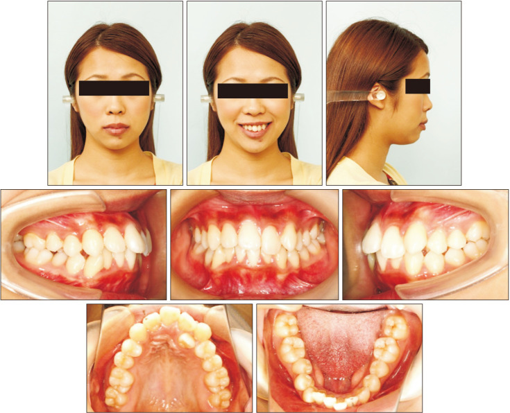
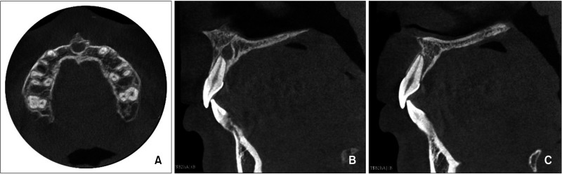
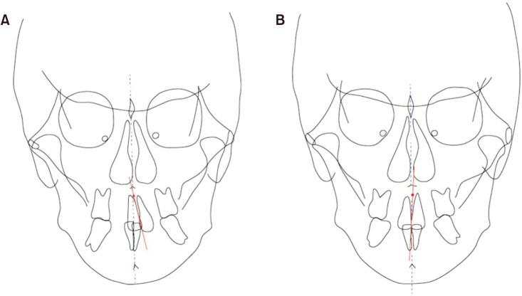
Root resorption can be caused by several factors, including contact with the cortical bone. Here we report a case involving a 21-year-old female with Angle Class II, division 1 malocclusion who exhibited significant root resorption in the maxillary right central incisor after orthodontic treatment. The patient presented with significant left-sided deviation of the maxillary incisors due to lingual dislocation of the left lateral incisor and a Class II molar relationship. Cephalometric analysis demonstrated a Class I skeletal relationship (A pointnasion-B point, 2.5°) and proclined maxillary anterior teeth (upper incisor to sella-nasion plane angle, 113.4°). The primary treatment objectives were the achievement of stable occlusion with midline agreement between the maxillary and mandibular dentitions and appropriate maxillary anterior tooth axes and molar relationship. A panoramic radiograph obtained after active treatment showed significant root resorption in the maxillary right central incisor; therefore, we performed cone-beam computed tomography, which confirmed root resorption along the cortical bone around the incisive canal. The findings from this case, where different degrees of root resorption were observed despite comparable degrees of orthodontic movement in the bilateral maxillary central incisors, suggest that the incisive canal could be an inducing factor for root resorption. However, further investigation is necessary to confirm this assumption.
期刊介绍:
The Korean Journal of Orthodontics (KJO) is an international, open access, peer reviewed journal published in January, March, May, July, September, and November each year. It was first launched in 1970 and, as the official scientific publication of Korean Association of Orthodontists, KJO aims to publish high quality clinical and scientific original research papers in all areas related to orthodontics and dentofacial orthopedics. Specifically, its interest focuses on evidence-based investigations of contemporary diagnostic procedures and treatment techniques, expanding to significant clinical reports of diverse treatment approaches.
The scope of KJO covers all areas of orthodontics and dentofacial orthopedics including successful diagnostic procedures and treatment planning, growth and development of the face and its clinical implications, appliance designs, biomechanics, TMJ disorders and adult treatment. Specifically, its latest interest focuses on skeletal anchorage devices, orthodontic appliance and biomaterials, 3 dimensional imaging techniques utilized for dentofacial diagnosis and treatment planning, and orthognathic surgery to correct skeletal disharmony in association of orthodontic treatment.


 分享
分享
 求助内容:
求助内容: 应助结果提醒方式:
 扫码关注我们
