{"title":"Localized Pretibial Varicose Vein Caused by an Intraosseous Venous Anomaly.","authors":"Sangwook Chun, Joung Woo Son, Jae-Wook Ryu","doi":"10.5090/kjtcs.2020.53.3.147","DOIUrl":null,"url":null,"abstract":"<p><p>A 36-year-old man presented to the hospital with protruding blood vessels in his left lower leg accompanied by cramping. An ultrasonographic examination of the leg revealed focal reflux without truncal vein reflux. During phlebectomy, the varix was found to be connected to the intraosseous vein through a tibial opening. Postoperative computed tomography and magnetic resonance imaging showed an osteolytic lesion in the tibial shaft and an intraosseous vascular anomaly. The patient was discharged without complications and scheduled for periodic follow-ups. This young man's varicose vein seemed to be from a tibial intraosseous vascular anomaly, which is extremely rare.</p>","PeriodicalId":38678,"journal":{"name":"Korean Journal of Thoracic and Cardiovascular Surgery","volume":"53 3","pages":"147-149"},"PeriodicalIF":0.0000,"publicationDate":"2020-06-05","publicationTypes":"Journal Article","fieldsOfStudy":null,"isOpenAccess":false,"openAccessPdf":"https://ftp.ncbi.nlm.nih.gov/pub/pmc/oa_pdf/53/15/KJTCV-53-147.PMC7287221.pdf","citationCount":"0","resultStr":null,"platform":"Semanticscholar","paperid":null,"PeriodicalName":"Korean Journal of Thoracic and Cardiovascular Surgery","FirstCategoryId":"1085","ListUrlMain":"https://doi.org/10.5090/kjtcs.2020.53.3.147","RegionNum":0,"RegionCategory":null,"ArticlePicture":[],"TitleCN":null,"AbstractTextCN":null,"PMCID":null,"EPubDate":"","PubModel":"","JCR":"Q3","JCRName":"Medicine","Score":null,"Total":0}
引用次数: 0
Abstract
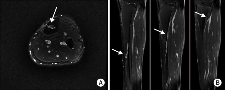
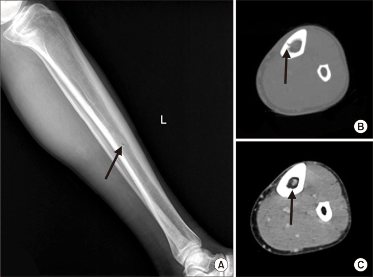
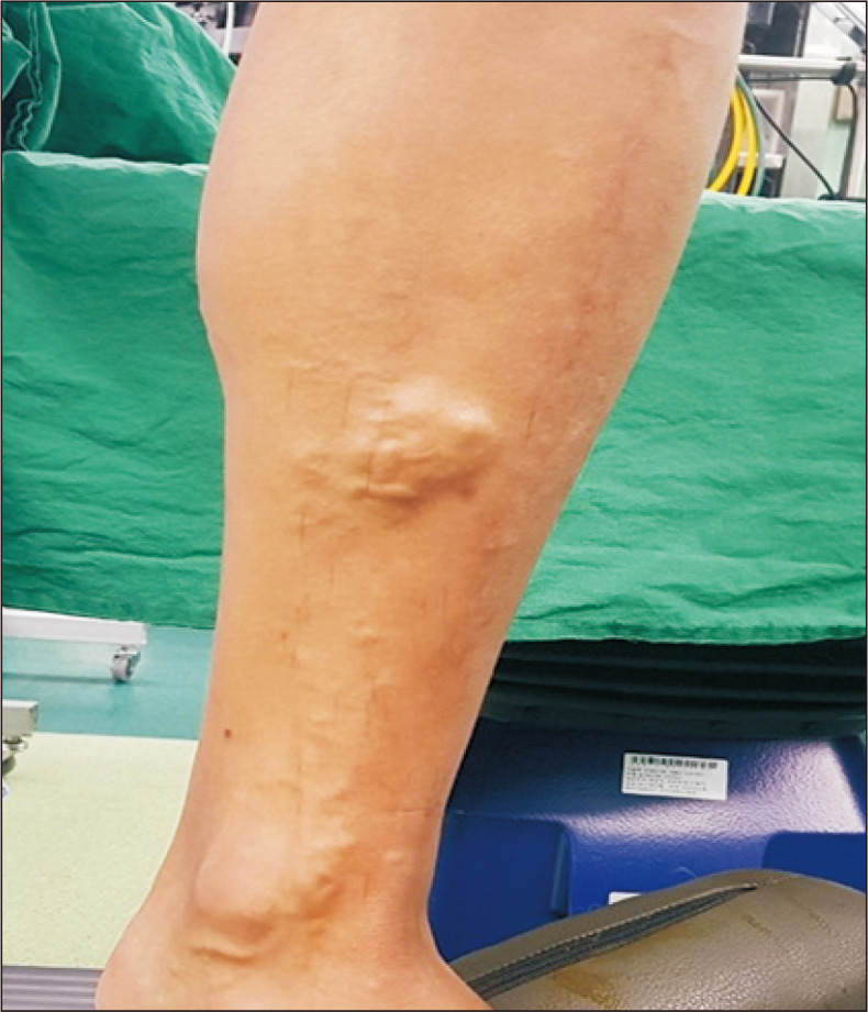
A 36-year-old man presented to the hospital with protruding blood vessels in his left lower leg accompanied by cramping. An ultrasonographic examination of the leg revealed focal reflux without truncal vein reflux. During phlebectomy, the varix was found to be connected to the intraosseous vein through a tibial opening. Postoperative computed tomography and magnetic resonance imaging showed an osteolytic lesion in the tibial shaft and an intraosseous vascular anomaly. The patient was discharged without complications and scheduled for periodic follow-ups. This young man's varicose vein seemed to be from a tibial intraosseous vascular anomaly, which is extremely rare.


 分享
分享
 求助内容:
求助内容: 应助结果提醒方式:
 扫码关注我们
