Amanda Parkes, Elizabeth Urquiola, Priya Bhosale, Heather Lin, Kelsey Watson, Wei-Lien Wang, Barry Feig, Keila Torres, Christina L Roland, Anthony P Conley, Maria Zarzour, J Andrew Livingston, Ravin Ratan, Joseph Ludwig, Dejka M Araujo, Vinod Ravi, Robert S Benjamin, Shreyaskumar Patel, Neeta Somaiah
{"title":"PET/CT Imaging as a Diagnostic Tool in Distinguishing Well-Differentiated versus Dedifferentiated Liposarcoma.","authors":"Amanda Parkes, Elizabeth Urquiola, Priya Bhosale, Heather Lin, Kelsey Watson, Wei-Lien Wang, Barry Feig, Keila Torres, Christina L Roland, Anthony P Conley, Maria Zarzour, J Andrew Livingston, Ravin Ratan, Joseph Ludwig, Dejka M Araujo, Vinod Ravi, Robert S Benjamin, Shreyaskumar Patel, Neeta Somaiah","doi":"10.1155/2020/8363986","DOIUrl":null,"url":null,"abstract":"<p><p>Distinguishing well-differentiated liposarcoma (WDLPS) from dedifferentiated liposarcoma (DDLPS) is essential given distinct treatment paradigms and chemosensitivity. Percutaneous biopsy has a low sensitivity for detecting DDLPS. We sought to identify the diagnostic utility of positron emission tomography/computed tomography (PET/CT) in identifying WDLPS versus DDLPS. An independent radiologist reviewed PET/CT images to identify target lesions and determine the maximum standardized uptake value (SUVmax). An independent pathologist review confirmed WDLPS or DDLPS histology. A binary cutoff point of SUVmax was identified using a classification and regression trees (CART) algorithm. We identified 20 patients with WDLPS or DDLPS with 26 PET/CTs performed for separate recurrences that were followed by surgical sampling. Of the 26 records, 12 were DDLPS (46%) and 14 were WDLPS (54%). Patients with DDLPS had significantly higher SUVmax than those with WDLPS (<i>p</i> value = 0.0035). A SUVmax of 4 was identified as the cutoff point. Using this cutoff, the sensitivity of SUVmax identifying a case as DDLPS was 83.3% (95% CI: 51.6%, 97.9%) and the specificity was 85.7% (95% CI: 57.2%, 98.2%). PET/CT is a sensitive and specific diagnostic tool to identify the presence of dedifferentiation within the tumor.</p>","PeriodicalId":21431,"journal":{"name":"Sarcoma","volume":"2020 ","pages":"8363986"},"PeriodicalIF":0.0000,"publicationDate":"2020-05-29","publicationTypes":"Journal Article","fieldsOfStudy":null,"isOpenAccess":false,"openAccessPdf":"https://sci-hub-pdf.com/10.1155/2020/8363986","citationCount":"15","resultStr":null,"platform":"Semanticscholar","paperid":null,"PeriodicalName":"Sarcoma","FirstCategoryId":"1085","ListUrlMain":"https://doi.org/10.1155/2020/8363986","RegionNum":0,"RegionCategory":null,"ArticlePicture":[],"TitleCN":null,"AbstractTextCN":null,"PMCID":null,"EPubDate":"2020/1/1 0:00:00","PubModel":"eCollection","JCR":"Q2","JCRName":"Medicine","Score":null,"Total":0}
引用次数: 15
Abstract
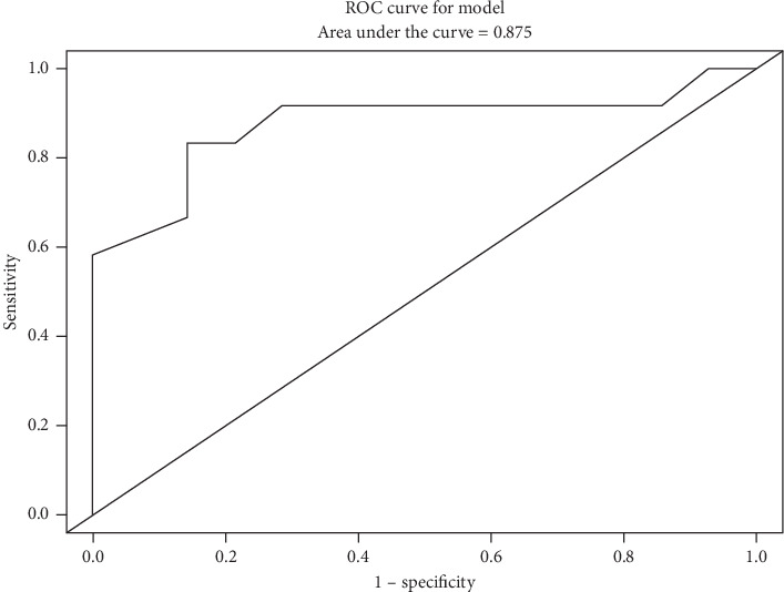
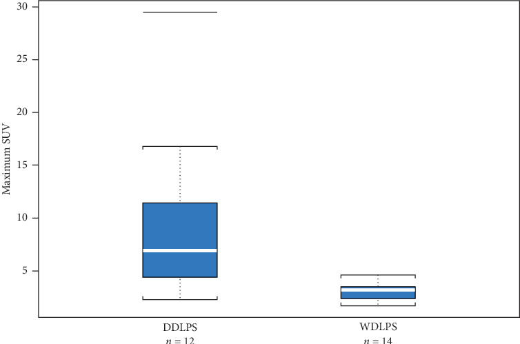
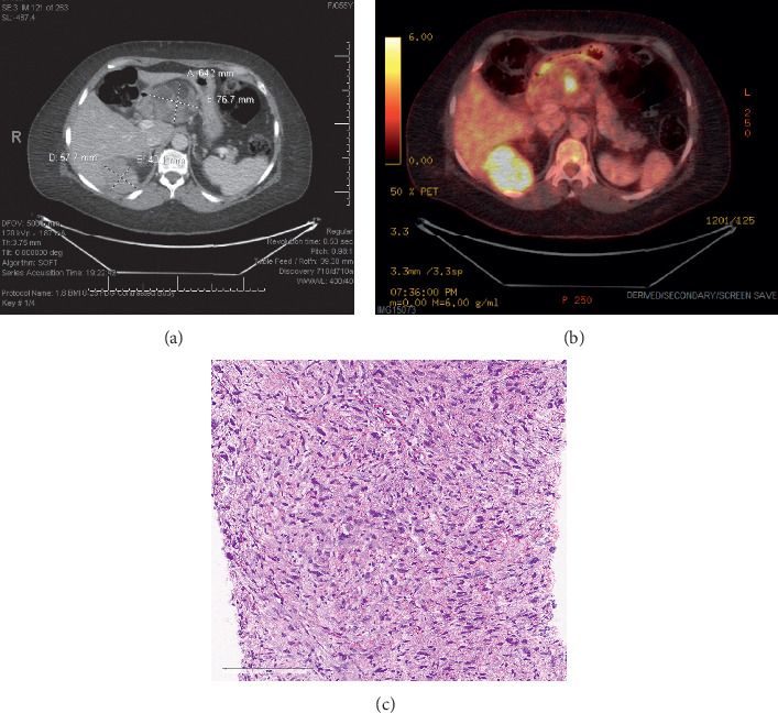
Distinguishing well-differentiated liposarcoma (WDLPS) from dedifferentiated liposarcoma (DDLPS) is essential given distinct treatment paradigms and chemosensitivity. Percutaneous biopsy has a low sensitivity for detecting DDLPS. We sought to identify the diagnostic utility of positron emission tomography/computed tomography (PET/CT) in identifying WDLPS versus DDLPS. An independent radiologist reviewed PET/CT images to identify target lesions and determine the maximum standardized uptake value (SUVmax). An independent pathologist review confirmed WDLPS or DDLPS histology. A binary cutoff point of SUVmax was identified using a classification and regression trees (CART) algorithm. We identified 20 patients with WDLPS or DDLPS with 26 PET/CTs performed for separate recurrences that were followed by surgical sampling. Of the 26 records, 12 were DDLPS (46%) and 14 were WDLPS (54%). Patients with DDLPS had significantly higher SUVmax than those with WDLPS (p value = 0.0035). A SUVmax of 4 was identified as the cutoff point. Using this cutoff, the sensitivity of SUVmax identifying a case as DDLPS was 83.3% (95% CI: 51.6%, 97.9%) and the specificity was 85.7% (95% CI: 57.2%, 98.2%). PET/CT is a sensitive and specific diagnostic tool to identify the presence of dedifferentiation within the tumor.
SarcomaMedicine-Radiology, Nuclear Medicine and Imaging
CiteScore
5.00
自引率
0.00%
发文量
15
审稿时长
14 weeks
期刊介绍:
Sarcoma is dedicated to publishing papers covering all aspects of connective tissue oncology research. It brings together work from scientists and clinicians carrying out a broad range of research in this field, including the basic sciences, molecular biology and pathology and the clinical sciences of epidemiology, surgery, radiotherapy and chemotherapy. High-quality papers concerning the entire range of bone and soft tissue sarcomas in both adults and children, including Kaposi"s sarcoma, are published as well as preclinical and animal studies. This journal provides a central forum for the description of advances in diagnosis, assessment and treatment of this rarely seen, but often mismanaged, group of patients.


 分享
分享
 求助内容:
求助内容: 应助结果提醒方式:
 扫码关注我们
