{"title":"Giant Protruding High-Grade Undifferentiated Pleomorphic Sarcoma Arising in a Keloid Scar on the Abdominal Wall.","authors":"Hideyuki Kinoshita, Toshinori Tsukanishi, Takeshi Ishii, Hiroto Kamoda, Yoko Hagiwara, Sumihisa Orita, Kazuhide Inage, Naoya Hirosawa, Seiji Ohtori, Tsukasa Yonemoto","doi":"10.1155/2020/4898965","DOIUrl":null,"url":null,"abstract":"<p><p>Undifferentiated pleomorphic sarcoma (UPS) is a high-grade, aggressive, soft tissue sarcoma that is often fatal. Although there are reports describing associations of sarcoma and skin lesions such as burns, radiation, and trauma, to our knowledge, UPS development in a keloid scar has not been reported. Herein, we present the case of a 76-year-old woman who had undergone surgery for endometrial cancer, 5 years before. She presented with a protruding lesion that was continuous to a keloid scar on the abdominal wall. The tumor appeared clinically malignant as it was protruding and doubled in size within three weeks, reaching approximately 6 × 6 × 2 cm. Since the tumor was diagnosed as UPS after pathological evaluation by needle biopsy, wide resection was performed. Intraoperatively, the tumor was apparently continuous to the keloid, protruding and pedunculated outside the body, and had not invaded the abdominal cavity. Histopathological examination of the resected tumor showed evidence of UPS and no suspicion of metastasis of endometrial cancer. No recurrence, metastases, or other complications were noted 6 months after surgery. The current case study reminds us that keloids may cause high-grade sarcoma such as UPS, and careful follow-up is required.</p>","PeriodicalId":9630,"journal":{"name":"Case Reports in Dermatological Medicine","volume":"2020 ","pages":"4898965"},"PeriodicalIF":0.0000,"publicationDate":"2020-07-24","publicationTypes":"Journal Article","fieldsOfStudy":null,"isOpenAccess":false,"openAccessPdf":"https://sci-hub-pdf.com/10.1155/2020/4898965","citationCount":"0","resultStr":null,"platform":"Semanticscholar","paperid":null,"PeriodicalName":"Case Reports in Dermatological Medicine","FirstCategoryId":"1085","ListUrlMain":"https://doi.org/10.1155/2020/4898965","RegionNum":0,"RegionCategory":null,"ArticlePicture":[],"TitleCN":null,"AbstractTextCN":null,"PMCID":null,"EPubDate":"2020/1/1 0:00:00","PubModel":"eCollection","JCR":"Q3","JCRName":"Medicine","Score":null,"Total":0}
引用次数: 0
Abstract
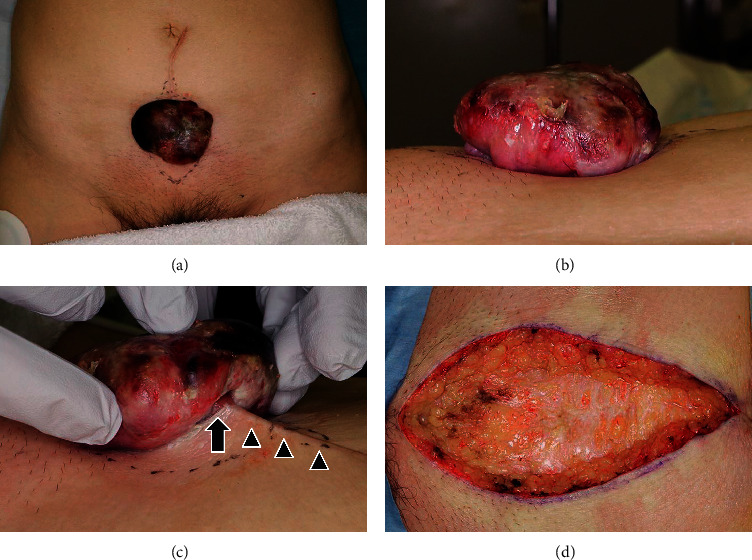
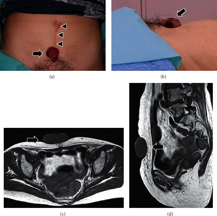
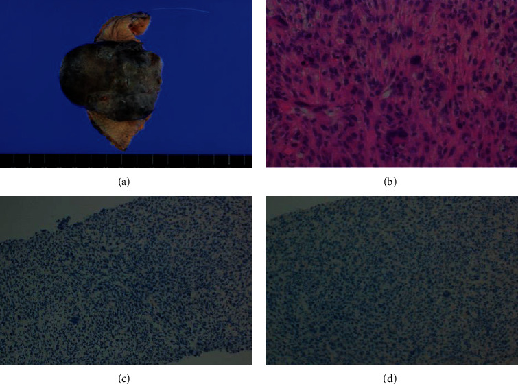
Undifferentiated pleomorphic sarcoma (UPS) is a high-grade, aggressive, soft tissue sarcoma that is often fatal. Although there are reports describing associations of sarcoma and skin lesions such as burns, radiation, and trauma, to our knowledge, UPS development in a keloid scar has not been reported. Herein, we present the case of a 76-year-old woman who had undergone surgery for endometrial cancer, 5 years before. She presented with a protruding lesion that was continuous to a keloid scar on the abdominal wall. The tumor appeared clinically malignant as it was protruding and doubled in size within three weeks, reaching approximately 6 × 6 × 2 cm. Since the tumor was diagnosed as UPS after pathological evaluation by needle biopsy, wide resection was performed. Intraoperatively, the tumor was apparently continuous to the keloid, protruding and pedunculated outside the body, and had not invaded the abdominal cavity. Histopathological examination of the resected tumor showed evidence of UPS and no suspicion of metastasis of endometrial cancer. No recurrence, metastases, or other complications were noted 6 months after surgery. The current case study reminds us that keloids may cause high-grade sarcoma such as UPS, and careful follow-up is required.


 分享
分享
 求助内容:
求助内容: 应助结果提醒方式:
 扫码关注我们
