{"title":"Fistulas between the Esophagus and Adjacent Vital Organs in Esophageal Cancer.","authors":"Sukki Cho","doi":"10.5090/kjtcs.2020.53.4.211","DOIUrl":null,"url":null,"abstract":"<p><p>Esophageal fistulas may occur in an advanced stage or as a potentially life-threatening complication of treatment. They can be divided into esophageal-respiratory and esophageal-aorta fistulas. The diagnosis is confirmed with fluoroscopy using dilute barium oral contrast, followed by thin-section computed tomography, which defines the precise location and extent of the fistula. Flexible esophagoscopy and bronchoscopy are required for confirmation and anatomic assessment of the suspected fistula and provide additional information for treatment planning. Contamination is traditionally controlled by surgical exclusion, along with a jejunal feeding tube. Currently, fully covered self-expanding metal stents are the primary treatment option.</p>","PeriodicalId":38678,"journal":{"name":"Korean Journal of Thoracic and Cardiovascular Surgery","volume":"53 4","pages":"211-216"},"PeriodicalIF":0.0000,"publicationDate":"2020-08-05","publicationTypes":"Journal Article","fieldsOfStudy":null,"isOpenAccess":false,"openAccessPdf":"https://ftp.ncbi.nlm.nih.gov/pub/pmc/oa_pdf/3f/16/KJTCV-53-211.PMC7409885.pdf","citationCount":"2","resultStr":null,"platform":"Semanticscholar","paperid":null,"PeriodicalName":"Korean Journal of Thoracic and Cardiovascular Surgery","FirstCategoryId":"1085","ListUrlMain":"https://doi.org/10.5090/kjtcs.2020.53.4.211","RegionNum":0,"RegionCategory":null,"ArticlePicture":[],"TitleCN":null,"AbstractTextCN":null,"PMCID":null,"EPubDate":"","PubModel":"","JCR":"Q3","JCRName":"Medicine","Score":null,"Total":0}
引用次数: 2
Abstract
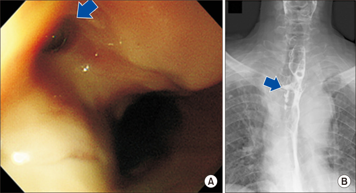
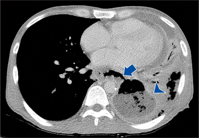
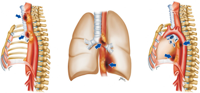
Esophageal fistulas may occur in an advanced stage or as a potentially life-threatening complication of treatment. They can be divided into esophageal-respiratory and esophageal-aorta fistulas. The diagnosis is confirmed with fluoroscopy using dilute barium oral contrast, followed by thin-section computed tomography, which defines the precise location and extent of the fistula. Flexible esophagoscopy and bronchoscopy are required for confirmation and anatomic assessment of the suspected fistula and provide additional information for treatment planning. Contamination is traditionally controlled by surgical exclusion, along with a jejunal feeding tube. Currently, fully covered self-expanding metal stents are the primary treatment option.


 分享
分享
 求助内容:
求助内容: 应助结果提醒方式:
 扫码关注我们
