Sandra Neumann, Elena G Milano, Chiara Bucciarelli-Ducci, Giovanni Biglino
{"title":"Imaging the carotid atherosclerotic plaque.","authors":"Sandra Neumann, Elena G Milano, Chiara Bucciarelli-Ducci, Giovanni Biglino","doi":"10.1530/VB-19-0010","DOIUrl":null,"url":null,"abstract":"<p><p>This mini review provides a concise overview of imaging techniques that are currently used to image the atheroscletoric plaque in the carotid artery <i>in vivo</i>. The main techniques include ultrasound imaging, X-ray imaging, magnetic resonance imaging and positron emission tomography imaging. Each technique has advantages and limitations and may be chosen depending on the availability, cost and clinical justification for its use. Common to all the imaging techniques presented here is the need for a skilled imaging professional to allow for high reliability and repeatability. While ultrasound-based imaging currently is regarded as a first line technique in clinical practice, the use of other techniques such as computed tomography angiography or magnetic resonance angiography need to be considered in the presence of significant stenosis with or without symptoms. Advancements in these two modalities, as well as in positron emission tomography imaging, are increasingly moving toward a better understanding of the risk-stratification and pre-interventional monitoring of patients at risk of plaque rupture as well as early identification of plaque development and better understanding of plaque composition (e.g. metabolic imaging).</p>","PeriodicalId":75294,"journal":{"name":"Vascular biology (Bristol, England)","volume":"1 1","pages":"H53-H58"},"PeriodicalIF":0.0000,"publicationDate":"2019-06-28","publicationTypes":"Journal Article","fieldsOfStudy":null,"isOpenAccess":false,"openAccessPdf":"https://www.ncbi.nlm.nih.gov/pmc/articles/PMC7439847/pdf/","citationCount":"0","resultStr":null,"platform":"Semanticscholar","paperid":null,"PeriodicalName":"Vascular biology (Bristol, England)","FirstCategoryId":"1085","ListUrlMain":"https://doi.org/10.1530/VB-19-0010","RegionNum":0,"RegionCategory":null,"ArticlePicture":[],"TitleCN":null,"AbstractTextCN":null,"PMCID":null,"EPubDate":"2019/1/1 0:00:00","PubModel":"eCollection","JCR":"","JCRName":"","Score":null,"Total":0}
引用次数: 0
Abstract
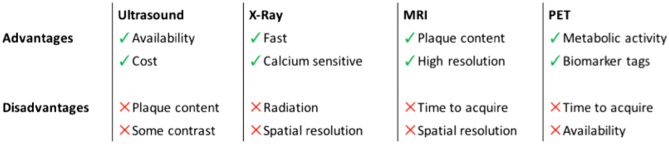
This mini review provides a concise overview of imaging techniques that are currently used to image the atheroscletoric plaque in the carotid artery in vivo. The main techniques include ultrasound imaging, X-ray imaging, magnetic resonance imaging and positron emission tomography imaging. Each technique has advantages and limitations and may be chosen depending on the availability, cost and clinical justification for its use. Common to all the imaging techniques presented here is the need for a skilled imaging professional to allow for high reliability and repeatability. While ultrasound-based imaging currently is regarded as a first line technique in clinical practice, the use of other techniques such as computed tomography angiography or magnetic resonance angiography need to be considered in the presence of significant stenosis with or without symptoms. Advancements in these two modalities, as well as in positron emission tomography imaging, are increasingly moving toward a better understanding of the risk-stratification and pre-interventional monitoring of patients at risk of plaque rupture as well as early identification of plaque development and better understanding of plaque composition (e.g. metabolic imaging).


 分享
分享

 求助内容:
求助内容: 应助结果提醒方式:
 扫码关注我们
