Seung Hwan Song, Chong Hoon Kim, Duk Hwan Moon, Sungsoo Lee
{"title":"Usefulness of 3-Dimensional Body Surface Scanning in the Evaluation of Patients with Pectus Carinatum.","authors":"Seung Hwan Song, Chong Hoon Kim, Duk Hwan Moon, Sungsoo Lee","doi":"10.5090/kjtcs.20.042","DOIUrl":null,"url":null,"abstract":"<p><strong>Background: </strong>Radiographic modalities have been commonly used to evaluate pectus carinatum (PC), and compressive orthotic bracing is the most widely accepted treatment method. The aim of this study was to determine the efficacy of 3-dimensional (3D) body surface scanning as an alternative modality for the evaluation of PC.</p><p><strong>Methods: </strong>The medical records of 63 patients with PC who were treated with compressive orthotic bracing therapy between July 2017 and February 2019 were retrospectively analyzed. Using both 2-view chest radiography (posteroanterior and lateral view) and 3D body scanning, the height of maximal protrusion of the chest wall was measured both before and after 2 weeks of bracing therapy. The difference between the pre- and post-treatment measurements was calculated for both modalities, and these differences were compared and analyzed.</p><p><strong>Results: </strong>Based on the comparison between the pre- and post-treatment radiographs, bracing therapy produced favorable outcomes in all patients (p<0.001). The measurements obtained via 3D scanning were strongly correlated with those obtained via chest radiography (r=0.60).</p><p><strong>Conclusion: </strong>Based on the findings of this study, 3D body surface scanning appears to be an effective, radiation-free, and simple method for the post-treatment follow-up evaluation of PC, and thus can be considered an alternative to radiography.</p>","PeriodicalId":38678,"journal":{"name":"Korean Journal of Thoracic and Cardiovascular Surgery","volume":"53 5","pages":"301-305"},"PeriodicalIF":0.0000,"publicationDate":"2020-10-05","publicationTypes":"Journal Article","fieldsOfStudy":null,"isOpenAccess":false,"openAccessPdf":"https://ftp.ncbi.nlm.nih.gov/pub/pmc/oa_pdf/03/9c/KJTCV-53-301.PMC7553828.pdf","citationCount":"2","resultStr":null,"platform":"Semanticscholar","paperid":null,"PeriodicalName":"Korean Journal of Thoracic and Cardiovascular Surgery","FirstCategoryId":"1085","ListUrlMain":"https://doi.org/10.5090/kjtcs.20.042","RegionNum":0,"RegionCategory":null,"ArticlePicture":[],"TitleCN":null,"AbstractTextCN":null,"PMCID":null,"EPubDate":"","PubModel":"","JCR":"Q3","JCRName":"Medicine","Score":null,"Total":0}
引用次数: 2
Abstract
Background: Radiographic modalities have been commonly used to evaluate pectus carinatum (PC), and compressive orthotic bracing is the most widely accepted treatment method. The aim of this study was to determine the efficacy of 3-dimensional (3D) body surface scanning as an alternative modality for the evaluation of PC.
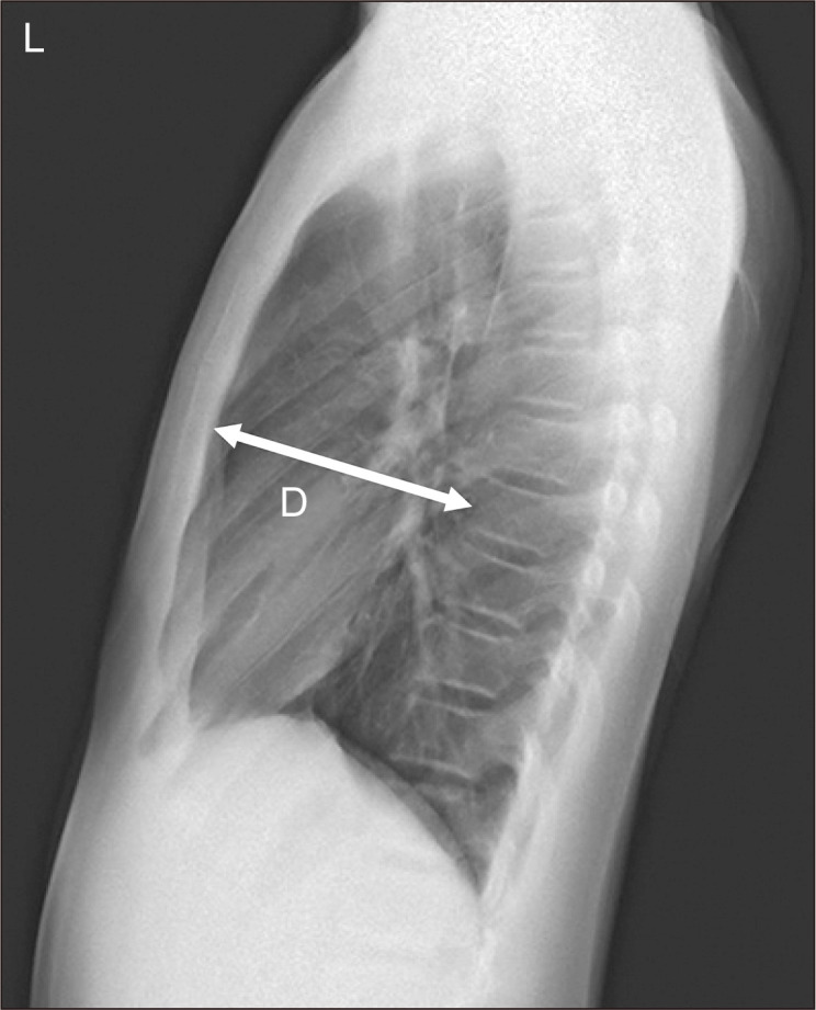
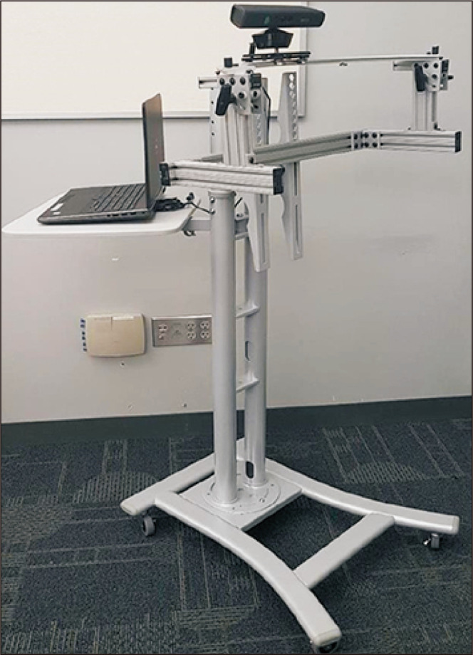
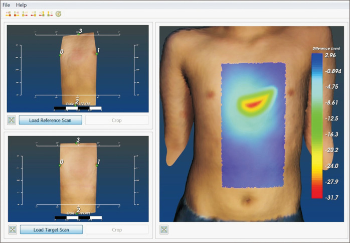
Methods: The medical records of 63 patients with PC who were treated with compressive orthotic bracing therapy between July 2017 and February 2019 were retrospectively analyzed. Using both 2-view chest radiography (posteroanterior and lateral view) and 3D body scanning, the height of maximal protrusion of the chest wall was measured both before and after 2 weeks of bracing therapy. The difference between the pre- and post-treatment measurements was calculated for both modalities, and these differences were compared and analyzed.
Results: Based on the comparison between the pre- and post-treatment radiographs, bracing therapy produced favorable outcomes in all patients (p<0.001). The measurements obtained via 3D scanning were strongly correlated with those obtained via chest radiography (r=0.60).
Conclusion: Based on the findings of this study, 3D body surface scanning appears to be an effective, radiation-free, and simple method for the post-treatment follow-up evaluation of PC, and thus can be considered an alternative to radiography.


 分享
分享
 求助内容:
求助内容: 应助结果提醒方式:
 扫码关注我们
