Andrew Spiro, Aqueel Usman, Asif Ajmal, Thanh D Hoang, Mohamed K M Shakir
{"title":"Asymptomatic and Biochemically Silent Pheochromocytoma with Characteristic Findings on Imaging.","authors":"Andrew Spiro, Aqueel Usman, Asif Ajmal, Thanh D Hoang, Mohamed K M Shakir","doi":"10.1155/2020/8847261","DOIUrl":null,"url":null,"abstract":"<p><p>Pheochromocytomas are tumors that originate from the chromaffin tissue of the adrenal medulla and commonly produce catecholamines. The diagnosis is typically established by the measurement of catecholamines or their metabolites in urine or plasma, and tumors are localized with the use of radiographic and scintigraphic studies. Pheochromocytomas can occur in asymptomatic patients, and the preferred treatment is surgical removal of the tumor. We report a 48-year-old male with a left adrenal incidentaloma, which progressively increased in size from 1.1 cm to 2.6 cm over a 4-year period, as measured by an adrenal computed tomography (CT) scan. Throughout his entire course of treatment, he was asymptomatic with normal blood pressure readings. His biochemical screening was unremarkable for the first three years of tumor surveillance. Follow-up imaging, including CT and MRI, showed findings suspicious for pheochromocytoma, and the diagnosis was ultimately made with the combination of imaging and laboratory studies. He underwent laparoscopic resection of the adrenal mass with confirmation of pheochromocytoma on histology. This case illustrates how CT and MRI findings can alert providers to the presence of a pheochromocytoma, even in an asymptomatic, biochemically negative patient.</p>","PeriodicalId":9621,"journal":{"name":"Case Reports in Endocrinology","volume":"2020 ","pages":"8847261"},"PeriodicalIF":0.9000,"publicationDate":"2020-09-26","publicationTypes":"Journal Article","fieldsOfStudy":null,"isOpenAccess":false,"openAccessPdf":"https://sci-hub-pdf.com/10.1155/2020/8847261","citationCount":"5","resultStr":null,"platform":"Semanticscholar","paperid":null,"PeriodicalName":"Case Reports in Endocrinology","FirstCategoryId":"1085","ListUrlMain":"https://doi.org/10.1155/2020/8847261","RegionNum":0,"RegionCategory":null,"ArticlePicture":[],"TitleCN":null,"AbstractTextCN":null,"PMCID":null,"EPubDate":"2020/1/1 0:00:00","PubModel":"eCollection","JCR":"Q4","JCRName":"ENDOCRINOLOGY & METABOLISM","Score":null,"Total":0}
引用次数: 5
Abstract
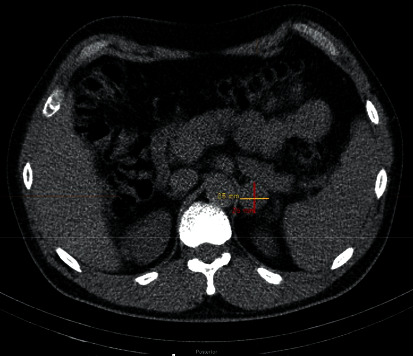
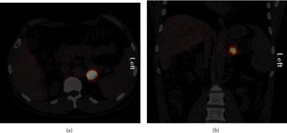
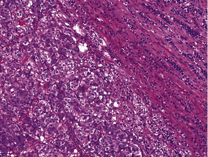
Pheochromocytomas are tumors that originate from the chromaffin tissue of the adrenal medulla and commonly produce catecholamines. The diagnosis is typically established by the measurement of catecholamines or their metabolites in urine or plasma, and tumors are localized with the use of radiographic and scintigraphic studies. Pheochromocytomas can occur in asymptomatic patients, and the preferred treatment is surgical removal of the tumor. We report a 48-year-old male with a left adrenal incidentaloma, which progressively increased in size from 1.1 cm to 2.6 cm over a 4-year period, as measured by an adrenal computed tomography (CT) scan. Throughout his entire course of treatment, he was asymptomatic with normal blood pressure readings. His biochemical screening was unremarkable for the first three years of tumor surveillance. Follow-up imaging, including CT and MRI, showed findings suspicious for pheochromocytoma, and the diagnosis was ultimately made with the combination of imaging and laboratory studies. He underwent laparoscopic resection of the adrenal mass with confirmation of pheochromocytoma on histology. This case illustrates how CT and MRI findings can alert providers to the presence of a pheochromocytoma, even in an asymptomatic, biochemically negative patient.


 分享
分享
 求助内容:
求助内容: 应助结果提醒方式:
 扫码关注我们
