Courtney Lawhn-Heath, Sue S Yom, Chienying Liu, Javier E Villanueva-Meyer, Maya Aslam, Raven Smith, Manpreet Narwal, Roxanna Juarez, Spencer C Behr, Miguel Hernandez Pampaloni, Jason W Chan, Christine M Glastonbury, Thomas A Hope, Robert R Flavell
{"title":"Gallium-68 prostate-specific membrane antigen ([<sup>68</sup>Ga]Ga-PSMA-11) PET for imaging of thyroid cancer: a feasibility study.","authors":"Courtney Lawhn-Heath, Sue S Yom, Chienying Liu, Javier E Villanueva-Meyer, Maya Aslam, Raven Smith, Manpreet Narwal, Roxanna Juarez, Spencer C Behr, Miguel Hernandez Pampaloni, Jason W Chan, Christine M Glastonbury, Thomas A Hope, Robert R Flavell","doi":"10.1186/s13550-020-00720-3","DOIUrl":null,"url":null,"abstract":"<p><strong>Background: </strong>Prostate-specific membrane antigen (PSMA) is expressed in the microvasculature of thyroid cancer. This suggests the potential use of PSMA as a diagnostic agent in patients with aggressive forms of thyroid cancer. The purpose of the current study was to determine the feasibility and utility of [<sup>68</sup>Ga]Ga-PSMA-11 PET/MRI in thyroid cancer patients.</p><p><strong>Methods: </strong>Eligible patients for this prospective pilot study were adults with a history of pathology-proven thyroid cancer who had abnormal radiotracer uptake on an 2-[<sup>18</sup>F]FDG PET and/or <sup>131</sup>I scintigraphy performed in the 12 months prior to study enrollment. Patients underwent a [<sup>68</sup>Ga]Ga-PSMA-11 PET/MRI, and comparison was made to the prior qualifying 2-[<sup>18</sup>F]FDG PET CT/MRI for lesion location and relative intensity.</p><p><strong>Results: </strong>Twelve patients underwent [<sup>68</sup>Ga]Ga-PSMA-11 PET/MRI, one of which was excluded from analysis due to debulking surgery prior to the PSMA PET. Of the remaining patients, 7/11 had differentiated disease (3 papillary, 2 follicular, 2 Hurthle cell) and 4/11 had dedifferentiated disease (2 poorly differentiated papillary, 2 anaplastic). Out of 43 lesions, 41 were visually 2-[<sup>18</sup>F]FDG positive (uptake greater than background, detection rate 95.3%) and 28 were PSMA positive (uptake greater than background, detection rate 65.1%). Uptake was heterogeneous between patients, and in some cases within patients. 3/11 patients (1 poorly differentiated papillary, 2 follicular) had PSMA uptake which was greater than FDG uptake. For the remaining 8 patients, 2-[<sup>18</sup>F]FDG uptake was greater than PSMA. Using one eligibility guideline in the prostate cancer literature for PSMA radioligand therapy (RLT), 8/11 could be considered eligible for possible future PSMA RLT. This was not predictable based on thyroid cancer subtype.</p><p><strong>Conclusions: </strong>[<sup>68</sup>Ga]Ga-PSMA-11 PET demonstrated lower detection rate when compared to 2-[<sup>18</sup>F]FDG PET for thyroid cancer lesion visualization. Thyroid cancer subtype alone may not be sufficient to predict PSMA uptake, and radiotracer uptake may vary between patients and even within patients.</p>","PeriodicalId":11611,"journal":{"name":"EJNMMI Research","volume":" ","pages":"128"},"PeriodicalIF":3.1000,"publicationDate":"2020-10-22","publicationTypes":"Journal Article","fieldsOfStudy":null,"isOpenAccess":false,"openAccessPdf":"https://sci-hub-pdf.com/10.1186/s13550-020-00720-3","citationCount":"16","resultStr":null,"platform":"Semanticscholar","paperid":null,"PeriodicalName":"EJNMMI Research","FirstCategoryId":"3","ListUrlMain":"https://doi.org/10.1186/s13550-020-00720-3","RegionNum":3,"RegionCategory":"医学","ArticlePicture":[],"TitleCN":null,"AbstractTextCN":null,"PMCID":null,"EPubDate":"","PubModel":"","JCR":"Q1","JCRName":"RADIOLOGY, NUCLEAR MEDICINE & MEDICAL IMAGING","Score":null,"Total":0}
引用次数: 16
Abstract
Background: Prostate-specific membrane antigen (PSMA) is expressed in the microvasculature of thyroid cancer. This suggests the potential use of PSMA as a diagnostic agent in patients with aggressive forms of thyroid cancer. The purpose of the current study was to determine the feasibility and utility of [68Ga]Ga-PSMA-11 PET/MRI in thyroid cancer patients.
Methods: Eligible patients for this prospective pilot study were adults with a history of pathology-proven thyroid cancer who had abnormal radiotracer uptake on an 2-[18F]FDG PET and/or 131I scintigraphy performed in the 12 months prior to study enrollment. Patients underwent a [68Ga]Ga-PSMA-11 PET/MRI, and comparison was made to the prior qualifying 2-[18F]FDG PET CT/MRI for lesion location and relative intensity.
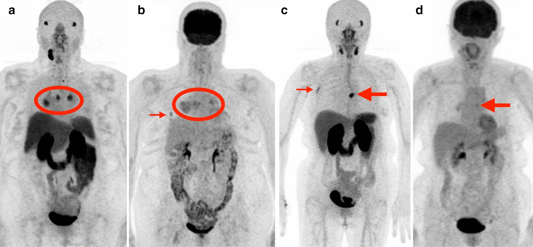
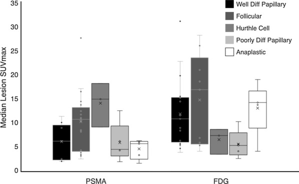
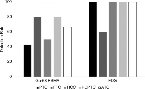
Results: Twelve patients underwent [68Ga]Ga-PSMA-11 PET/MRI, one of which was excluded from analysis due to debulking surgery prior to the PSMA PET. Of the remaining patients, 7/11 had differentiated disease (3 papillary, 2 follicular, 2 Hurthle cell) and 4/11 had dedifferentiated disease (2 poorly differentiated papillary, 2 anaplastic). Out of 43 lesions, 41 were visually 2-[18F]FDG positive (uptake greater than background, detection rate 95.3%) and 28 were PSMA positive (uptake greater than background, detection rate 65.1%). Uptake was heterogeneous between patients, and in some cases within patients. 3/11 patients (1 poorly differentiated papillary, 2 follicular) had PSMA uptake which was greater than FDG uptake. For the remaining 8 patients, 2-[18F]FDG uptake was greater than PSMA. Using one eligibility guideline in the prostate cancer literature for PSMA radioligand therapy (RLT), 8/11 could be considered eligible for possible future PSMA RLT. This was not predictable based on thyroid cancer subtype.
Conclusions: [68Ga]Ga-PSMA-11 PET demonstrated lower detection rate when compared to 2-[18F]FDG PET for thyroid cancer lesion visualization. Thyroid cancer subtype alone may not be sufficient to predict PSMA uptake, and radiotracer uptake may vary between patients and even within patients.
EJNMMI ResearchRADIOLOGY, NUCLEAR MEDICINE & MEDICAL IMAGING&nb-
CiteScore
5.90
自引率
3.10%
发文量
72
审稿时长
13 weeks
期刊介绍:
EJNMMI Research publishes new basic, translational and clinical research in the field of nuclear medicine and molecular imaging. Regular features include original research articles, rapid communication of preliminary data on innovative research, interesting case reports, editorials, and letters to the editor. Educational articles on basic sciences, fundamental aspects and controversy related to pre-clinical and clinical research or ethical aspects of research are also welcome. Timely reviews provide updates on current applications, issues in imaging research and translational aspects of nuclear medicine and molecular imaging technologies.
The main emphasis is placed on the development of targeted imaging with radiopharmaceuticals within the broader context of molecular probes to enhance understanding and characterisation of the complex biological processes underlying disease and to develop, test and guide new treatment modalities, including radionuclide therapy.


 分享
分享
 求助内容:
求助内容: 应助结果提醒方式:
 扫码关注我们
