Joshua Wei Liang Yip, Han Loh, Chuong Bui, Veronica Chi Ken Wong, Robert Mansberg
{"title":"False-Positive <sup>18</sup>F-FDG PET/CT Uptake in Unilateral Lactation.","authors":"Joshua Wei Liang Yip, Han Loh, Chuong Bui, Veronica Chi Ken Wong, Robert Mansberg","doi":"10.1155/2020/8850052","DOIUrl":null,"url":null,"abstract":"<p><p>A 31-year-old woman (7 months postpartum and lactating) with multiple sclerotic bone lesions was referred for an <sup>18</sup>F-FDG PET/CT scan for characterization. The scan demonstrated unilateral diffuse intense FDG uptake corresponding to dense soft tissue in the right breast, likely related to secretory hyperplasia. On further questioning, it was made apparent that she had only been breastfeeding from the right breast. While the left breast also demonstrated dense soft tissue to a lesser degree, no significant FDG uptake was seen. The sclerotic bone lesions were not FDG avid, likely due to a separate non-FDG avid benign condition or bony metastases from a non-FDG avid primary malignancy. This was reinforced by the fact that subsequent investigations including serial bilateral breast ultrasound and percutaneous biopsy demonstrated no definite evidence of malignancy in the bilateral breasts. The histopathology findings of an open surgical biopsy of sclerotic lesions in the left posterior ilium were also nonspecific, favouring bone dysplasia with no evidence of malignancy.</p>","PeriodicalId":30326,"journal":{"name":"Case Reports in Radiology","volume":"2020 ","pages":"8850052"},"PeriodicalIF":0.0000,"publicationDate":"2020-11-28","publicationTypes":"Journal Article","fieldsOfStudy":null,"isOpenAccess":false,"openAccessPdf":"https://sci-hub-pdf.com/10.1155/2020/8850052","citationCount":"0","resultStr":null,"platform":"Semanticscholar","paperid":null,"PeriodicalName":"Case Reports in Radiology","FirstCategoryId":"1085","ListUrlMain":"https://doi.org/10.1155/2020/8850052","RegionNum":0,"RegionCategory":null,"ArticlePicture":[],"TitleCN":null,"AbstractTextCN":null,"PMCID":null,"EPubDate":"2020/1/1 0:00:00","PubModel":"eCollection","JCR":"","JCRName":"","Score":null,"Total":0}
引用次数: 0
Abstract
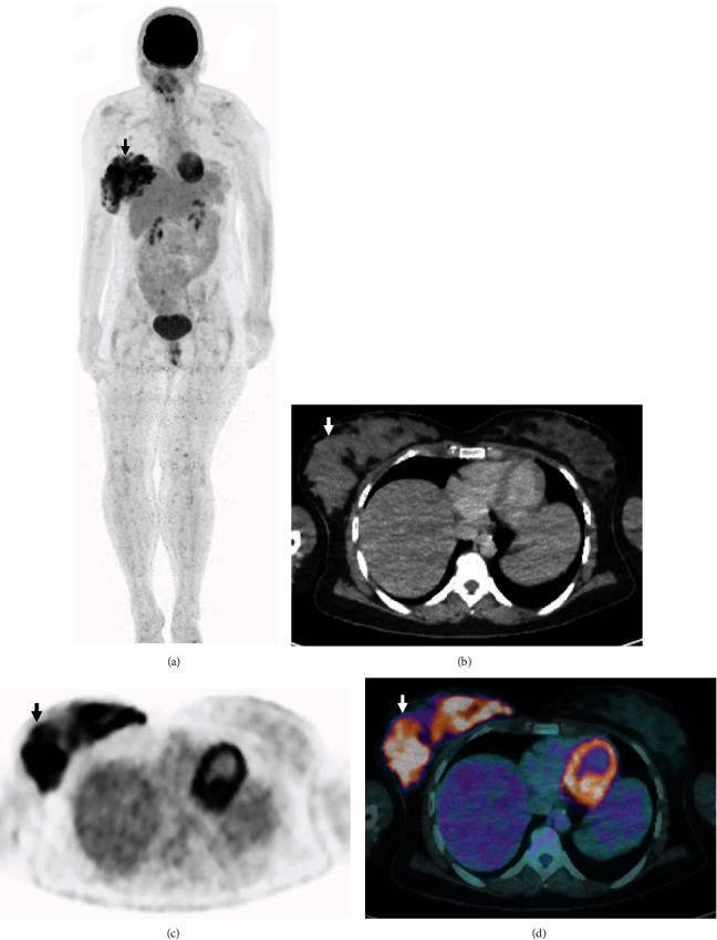
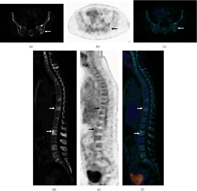
A 31-year-old woman (7 months postpartum and lactating) with multiple sclerotic bone lesions was referred for an 18F-FDG PET/CT scan for characterization. The scan demonstrated unilateral diffuse intense FDG uptake corresponding to dense soft tissue in the right breast, likely related to secretory hyperplasia. On further questioning, it was made apparent that she had only been breastfeeding from the right breast. While the left breast also demonstrated dense soft tissue to a lesser degree, no significant FDG uptake was seen. The sclerotic bone lesions were not FDG avid, likely due to a separate non-FDG avid benign condition or bony metastases from a non-FDG avid primary malignancy. This was reinforced by the fact that subsequent investigations including serial bilateral breast ultrasound and percutaneous biopsy demonstrated no definite evidence of malignancy in the bilateral breasts. The histopathology findings of an open surgical biopsy of sclerotic lesions in the left posterior ilium were also nonspecific, favouring bone dysplasia with no evidence of malignancy.


 分享
分享

 求助内容:
求助内容: 应助结果提醒方式:
 扫码关注我们
