{"title":"Correlation of Mast Cell and Angiogenesis in Oral Lichen Planus, Dysplasia (Leukoplakia), and Oral Squamous Cell Carcinoma.","authors":"Amutha Sundararajan, Rajmohan Muthusamy, Kumar Gopal Siva, Prasad Harikrishnan, Sri Chinthu Kenniyan Kumar, Selva Kumar Rathinasamy","doi":"10.5041/RMMJ.10438","DOIUrl":null,"url":null,"abstract":"<p><strong>Objective: </strong>The aim of this study was to compare and correlate mast cell density (MCD) and microvessel density (MVD) between normal oral mucosa, oral lichen planus, various grades of dysplasia, and oral squamous cell carcinoma (OSCC).</p><p><strong>Materials and methods: </strong>The study comprised a total of 75 samples, of which 65 were archival tissue blocks of histopathologically confirmed cases, which included 10 cases of oral lichen planus, 25 cases of dysplasia (mild [n=10], moderate [n=10], and severe [n=5]), and 30 cases of OSCC (well differentiated [n=10], moderately differentiated [n=10], and poorly differentiated [n=10]), and 10 samples of normal oral mucosa. All the sections were immunohistochemically stained with anti-CD34 and counterstained with toluidine blue stain. Mean MCD and MVD were determined and analyzed using ANOVA test and compared between the lesions using Tukey HSD test. Pearson's correlation coefficient test was used to correlate these two factors between various lesions.</p><p><strong>Results: </strong>Mean MCD and mean MVD were found to be increased in all the lesions compared to normal oral mucosa, and the values were statically significant. Overall, MCD and MVD showed a significant positive correlation (r=0.640).</p><p><strong>Conclusion: </strong>Increase of MCD and MVD and their positive correlation in all the lesions have emphasized their role in the pathogenesis and disease progression.</p>","PeriodicalId":46408,"journal":{"name":"Rambam Maimonides Medical Journal","volume":"12 2","pages":""},"PeriodicalIF":1.3000,"publicationDate":"2021-04-29","publicationTypes":"Journal Article","fieldsOfStudy":null,"isOpenAccess":false,"openAccessPdf":"https://www.ncbi.nlm.nih.gov/pmc/articles/PMC8092953/pdf/","citationCount":"6","resultStr":null,"platform":"Semanticscholar","paperid":null,"PeriodicalName":"Rambam Maimonides Medical Journal","FirstCategoryId":"1085","ListUrlMain":"https://doi.org/10.5041/RMMJ.10438","RegionNum":0,"RegionCategory":null,"ArticlePicture":[],"TitleCN":null,"AbstractTextCN":null,"PMCID":null,"EPubDate":"","PubModel":"","JCR":"Q2","JCRName":"MEDICINE, GENERAL & INTERNAL","Score":null,"Total":0}
引用次数: 6
Abstract
Objective: The aim of this study was to compare and correlate mast cell density (MCD) and microvessel density (MVD) between normal oral mucosa, oral lichen planus, various grades of dysplasia, and oral squamous cell carcinoma (OSCC).
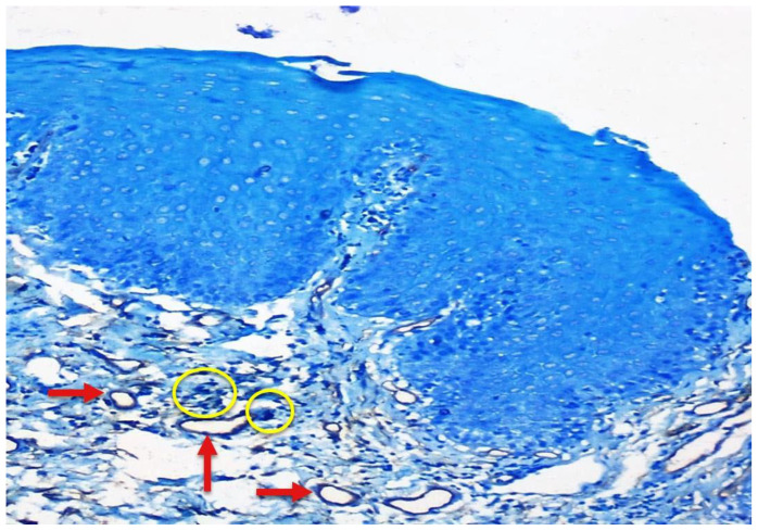
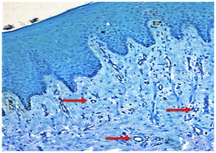
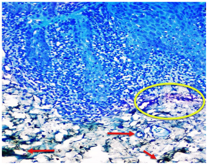
Materials and methods: The study comprised a total of 75 samples, of which 65 were archival tissue blocks of histopathologically confirmed cases, which included 10 cases of oral lichen planus, 25 cases of dysplasia (mild [n=10], moderate [n=10], and severe [n=5]), and 30 cases of OSCC (well differentiated [n=10], moderately differentiated [n=10], and poorly differentiated [n=10]), and 10 samples of normal oral mucosa. All the sections were immunohistochemically stained with anti-CD34 and counterstained with toluidine blue stain. Mean MCD and MVD were determined and analyzed using ANOVA test and compared between the lesions using Tukey HSD test. Pearson's correlation coefficient test was used to correlate these two factors between various lesions.
Results: Mean MCD and mean MVD were found to be increased in all the lesions compared to normal oral mucosa, and the values were statically significant. Overall, MCD and MVD showed a significant positive correlation (r=0.640).
Conclusion: Increase of MCD and MVD and their positive correlation in all the lesions have emphasized their role in the pathogenesis and disease progression.


 分享
分享
 求助内容:
求助内容: 应助结果提醒方式:
 扫码关注我们
