Klenam Dzefi-Tettey, Emmanuel Kobina Mesi Edzie, Philip Narteh Gorleku, Henry Kusodzi, Abdul Raman Asemah
{"title":"Asymptomatic Familial Multiple Cerebral Cavernous Malformation in a 73-Year-Old Woman.","authors":"Klenam Dzefi-Tettey, Emmanuel Kobina Mesi Edzie, Philip Narteh Gorleku, Henry Kusodzi, Abdul Raman Asemah","doi":"10.1155/2021/9974776","DOIUrl":null,"url":null,"abstract":"<p><p>Cerebral cavernous malformations (CCMs) are dilated blood vessels which can develop sporadically or in familial form and are the commonest malformations of blood vessels in the spinal cord and brain. The familial form is an autosomal dominant gene mutation disorder. This condition can be diagnosed with magnetic resonance imaging (MRI) and computed tomography (CT) scan, but the modality of choice is MRI because of its high sensitivity. We report a case of a 73-year-old woman with an asymptomatic multiple familial cerebral cavernous malformation (FCCM) which was previously misdiagnosed as multiple cerebral metastases on CT scan. A brain MRI performed correctly diagnosed her condition as FCCM based on the typical MRI appearances. In order not to misdiagnose brain lesions like CCM on CT scan, for cerebral metastases in resource-poor settings, radiologists must recommend advanced imaging modalities like MRI for further evaluation, thereby avoiding unnecessary invasive surgical biopsies.</p>","PeriodicalId":30326,"journal":{"name":"Case Reports in Radiology","volume":" ","pages":"9974776"},"PeriodicalIF":0.0000,"publicationDate":"2021-05-21","publicationTypes":"Journal Article","fieldsOfStudy":null,"isOpenAccess":false,"openAccessPdf":"https://www.ncbi.nlm.nih.gov/pmc/articles/PMC8163527/pdf/","citationCount":"0","resultStr":null,"platform":"Semanticscholar","paperid":null,"PeriodicalName":"Case Reports in Radiology","FirstCategoryId":"1085","ListUrlMain":"https://doi.org/10.1155/2021/9974776","RegionNum":0,"RegionCategory":null,"ArticlePicture":[],"TitleCN":null,"AbstractTextCN":null,"PMCID":null,"EPubDate":"2021/1/1 0:00:00","PubModel":"eCollection","JCR":"","JCRName":"","Score":null,"Total":0}
引用次数: 0
Abstract
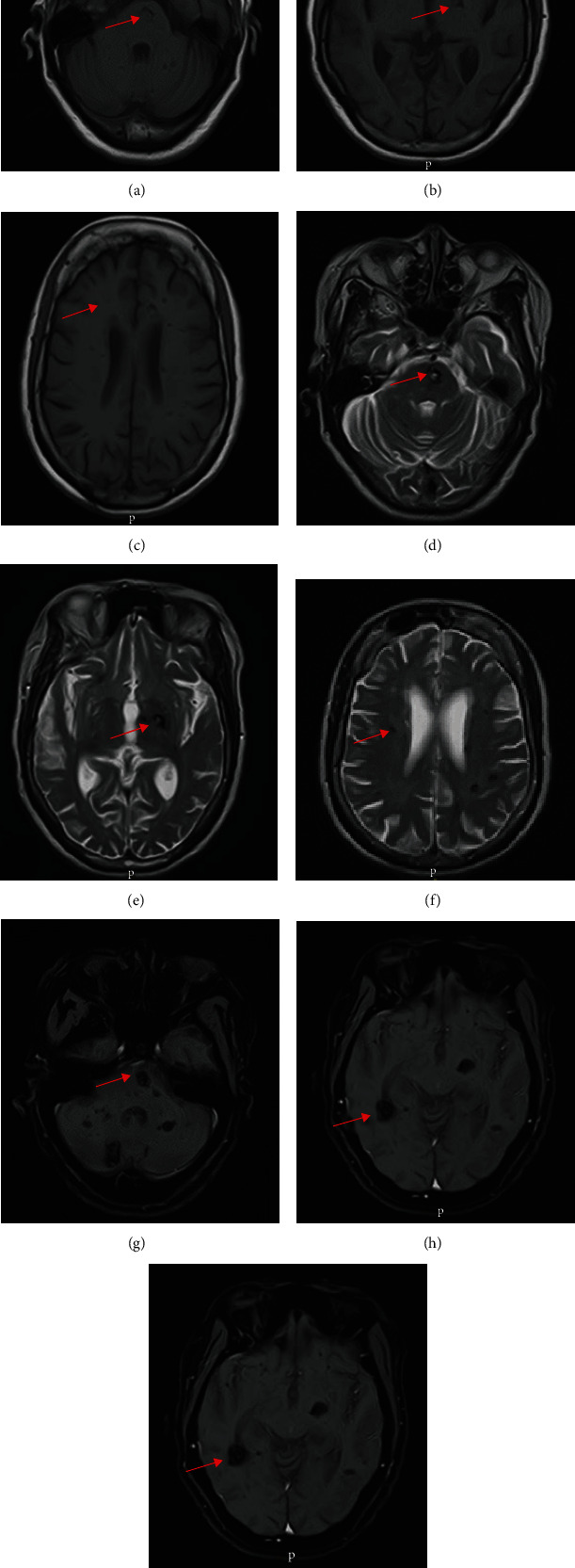
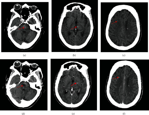
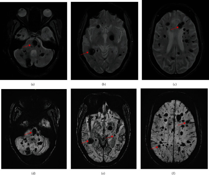
Cerebral cavernous malformations (CCMs) are dilated blood vessels which can develop sporadically or in familial form and are the commonest malformations of blood vessels in the spinal cord and brain. The familial form is an autosomal dominant gene mutation disorder. This condition can be diagnosed with magnetic resonance imaging (MRI) and computed tomography (CT) scan, but the modality of choice is MRI because of its high sensitivity. We report a case of a 73-year-old woman with an asymptomatic multiple familial cerebral cavernous malformation (FCCM) which was previously misdiagnosed as multiple cerebral metastases on CT scan. A brain MRI performed correctly diagnosed her condition as FCCM based on the typical MRI appearances. In order not to misdiagnose brain lesions like CCM on CT scan, for cerebral metastases in resource-poor settings, radiologists must recommend advanced imaging modalities like MRI for further evaluation, thereby avoiding unnecessary invasive surgical biopsies.


 分享
分享

 求助内容:
求助内容: 应助结果提醒方式:
 扫码关注我们
