{"title":"Spontaneous (Hashimoto-like) chronic lymphocytic thyroiditis in a rhesus macaque (<i>Macaca mulatta</i>).","authors":"Roland Plesker, Gudrun Hintereder","doi":"10.5194/pb-8-37-2021","DOIUrl":null,"url":null,"abstract":"<p><p>A case of a female, 10-year-old rhesus macaque (<i>Macaca mulatta</i>) with spontaneous chronic lymphocytic thyroiditis is presented. At necropsy, the thyroid gland was slightly enlarged, with up to 2 mm large, round, confluent, beige foci on the surface of both lobes. Histopathologic features resembled human Hashimoto's thyroiditis: multifocally, the interstitium was infiltrated by lymphocytes and variably sized lymphoid follicles. In the pituitary gland, there were increased numbers of large, basophilic cells throughout the adenohypophysis. Using a human electrochemiluminescence immunoassay (ECLIA), no autoantibodies against thyroglobulin, thyroid peroxidase, or thyroid-stimulating hormone receptor were detected.</p>","PeriodicalId":37245,"journal":{"name":"Primate Biology","volume":"8 1","pages":"37-42"},"PeriodicalIF":0.0000,"publicationDate":"2021-06-01","publicationTypes":"Journal Article","fieldsOfStudy":null,"isOpenAccess":false,"openAccessPdf":"https://www.ncbi.nlm.nih.gov/pmc/articles/PMC8182663/pdf/","citationCount":"2","resultStr":null,"platform":"Semanticscholar","paperid":null,"PeriodicalName":"Primate Biology","FirstCategoryId":"1085","ListUrlMain":"https://doi.org/10.5194/pb-8-37-2021","RegionNum":0,"RegionCategory":null,"ArticlePicture":[],"TitleCN":null,"AbstractTextCN":null,"PMCID":null,"EPubDate":"2021/1/1 0:00:00","PubModel":"eCollection","JCR":"Q2","JCRName":"Agricultural and Biological Sciences","Score":null,"Total":0}
引用次数: 2
Abstract
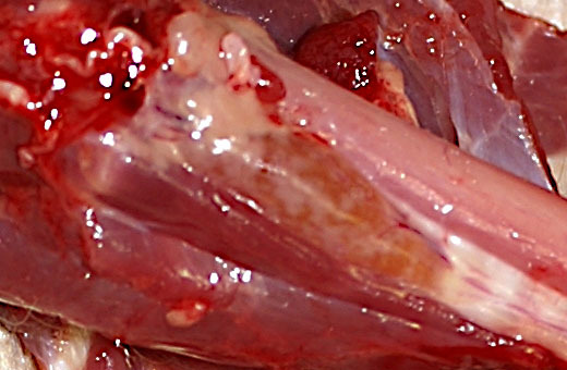
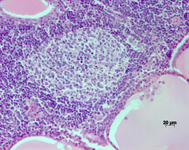
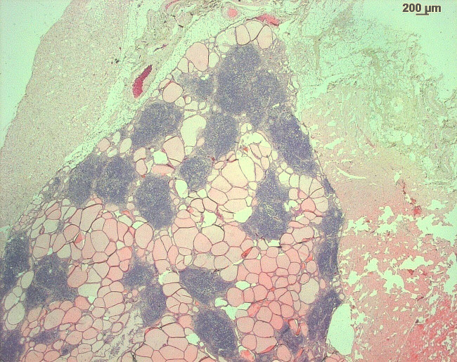
A case of a female, 10-year-old rhesus macaque (Macaca mulatta) with spontaneous chronic lymphocytic thyroiditis is presented. At necropsy, the thyroid gland was slightly enlarged, with up to 2 mm large, round, confluent, beige foci on the surface of both lobes. Histopathologic features resembled human Hashimoto's thyroiditis: multifocally, the interstitium was infiltrated by lymphocytes and variably sized lymphoid follicles. In the pituitary gland, there were increased numbers of large, basophilic cells throughout the adenohypophysis. Using a human electrochemiluminescence immunoassay (ECLIA), no autoantibodies against thyroglobulin, thyroid peroxidase, or thyroid-stimulating hormone receptor were detected.


 分享
分享
 求助内容:
求助内容: 应助结果提醒方式:
 扫码关注我们
