Chest CT Findings and Differential Diagnosis of Mycoplasma pneumoniae Pneumonia and Mycoplasma pneumoniae Combined with Streptococcal Pneumonia in Children.
{"title":"Chest CT Findings and Differential Diagnosis of <i>Mycoplasma pneumoniae</i> Pneumonia and <i>Mycoplasma pneumoniae</i> Combined with Streptococcal Pneumonia in Children.","authors":"Jing Wang, Chen Xia, Ashutosh Sharma, Gurjot Singh Gaba, Mohammad Shabaz","doi":"10.1155/2021/8085530","DOIUrl":null,"url":null,"abstract":"<p><strong>Background: </strong>In this day and age, 17% of children less than 5 years of age died of pneumonia; it is the common cause of children death. It is one of the main children respiratory infectious diseases, i.e., mycoplasma pneumonia (MP). The imaging examination can be adopted to quickly observe the morphology and scope of the pulmonary lesions and know the effect of disease treatment and subsequent changes in the disease in order to provide a basis for treatment. Therefore, the most commonly applied technology for detecting pneumonia in children is imaging technology, including chest X-ray and CT.</p><p><strong>Objectives: </strong>The main objective of the work is to investigate the chest computed tomography (CT) findings of children patients with <i>Mycoplasma pneumoniae</i> pneumonia (MPP) and MP combined with streptococcal pneumonia (SP). The mixed infection of MP and SP is very common clinically, and the diagnosis of this type of mixed pneumonia is a critical research topic faced by pediatric respiratory physicians. The comparison is done on the incidence of bronchial and pulmonary interstitial lesions, the degree of lymph node enlargement, the volume and depth of pleural effusion, and the location and morphology of the pulmonary lesions in the chest CT images of children patients from the two groups.</p><p><strong>Methods: </strong>There were comparisons on the incidence of bronchial and pulmonary interstitial lesions, the degree of lymph node enlargement, the volume and depth of pleural effusion, and the location and morphology of the pulmonary lesions in the chest CT images of children patients from the two groups. All the experiments are done in the MATLAB.</p><p><strong>Results: </strong>The results showed that the proportions of reticular shadow, ground glass shadow, bronchial inflation phase, tube wall thickening, and vascular bundle thickening on the CT images of children patients from the MPP group were dramatically higher than those of the MP + SP group (<i>P</i> < 0.05). The maximum transverse diameter of enlarged lymph node in children patients from the MPP group was obviously larger than the diameter of the MP + SP group (<i>P</i> < 0.05). The number of children patients with pleural effusion was 22 in the MP + SP group, which was greatly higher than the MPP group (<i>P</i> < 0.05).</p><p><strong>Conclusion: </strong>In conclusion, the chest CT images of children patients from the MPP group were mainly pulmonary interstitial changes. Furthermore, the alveolar inflammation could be observed on the CT images shown when children patients were combined with SP infection. The more obvious manifestations were that the flaky shadows appeared in the lungs, the pleural effusion became thicker, and the transverse diameters of enlarged lymph nodes were bigger.</p>","PeriodicalId":16017,"journal":{"name":"Journal of Healthcare Engineering","volume":" ","pages":"8085530"},"PeriodicalIF":0.0000,"publicationDate":"2021-06-14","publicationTypes":"Journal Article","fieldsOfStudy":null,"isOpenAccess":false,"openAccessPdf":"https://www.ncbi.nlm.nih.gov/pmc/articles/PMC8219438/pdf/","citationCount":"18","resultStr":null,"platform":"Semanticscholar","paperid":null,"PeriodicalName":"Journal of Healthcare Engineering","FirstCategoryId":"3","ListUrlMain":"https://doi.org/10.1155/2021/8085530","RegionNum":4,"RegionCategory":"医学","ArticlePicture":[],"TitleCN":null,"AbstractTextCN":null,"PMCID":null,"EPubDate":"2021/1/1 0:00:00","PubModel":"eCollection","JCR":"Q2","JCRName":"Medicine","Score":null,"Total":0}
引用次数: 18
Abstract
Background: In this day and age, 17% of children less than 5 years of age died of pneumonia; it is the common cause of children death. It is one of the main children respiratory infectious diseases, i.e., mycoplasma pneumonia (MP). The imaging examination can be adopted to quickly observe the morphology and scope of the pulmonary lesions and know the effect of disease treatment and subsequent changes in the disease in order to provide a basis for treatment. Therefore, the most commonly applied technology for detecting pneumonia in children is imaging technology, including chest X-ray and CT.
Objectives: The main objective of the work is to investigate the chest computed tomography (CT) findings of children patients with Mycoplasma pneumoniae pneumonia (MPP) and MP combined with streptococcal pneumonia (SP). The mixed infection of MP and SP is very common clinically, and the diagnosis of this type of mixed pneumonia is a critical research topic faced by pediatric respiratory physicians. The comparison is done on the incidence of bronchial and pulmonary interstitial lesions, the degree of lymph node enlargement, the volume and depth of pleural effusion, and the location and morphology of the pulmonary lesions in the chest CT images of children patients from the two groups.
Methods: There were comparisons on the incidence of bronchial and pulmonary interstitial lesions, the degree of lymph node enlargement, the volume and depth of pleural effusion, and the location and morphology of the pulmonary lesions in the chest CT images of children patients from the two groups. All the experiments are done in the MATLAB.
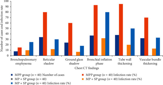
Results: The results showed that the proportions of reticular shadow, ground glass shadow, bronchial inflation phase, tube wall thickening, and vascular bundle thickening on the CT images of children patients from the MPP group were dramatically higher than those of the MP + SP group (P < 0.05). The maximum transverse diameter of enlarged lymph node in children patients from the MPP group was obviously larger than the diameter of the MP + SP group (P < 0.05). The number of children patients with pleural effusion was 22 in the MP + SP group, which was greatly higher than the MPP group (P < 0.05).
Conclusion: In conclusion, the chest CT images of children patients from the MPP group were mainly pulmonary interstitial changes. Furthermore, the alveolar inflammation could be observed on the CT images shown when children patients were combined with SP infection. The more obvious manifestations were that the flaky shadows appeared in the lungs, the pleural effusion became thicker, and the transverse diameters of enlarged lymph nodes were bigger.
期刊介绍:
The Journal of Healthcare Engineering is a peer-reviewed, Open Access journal publishing fundamental and applied research on all aspects of engineering involved in healthcare delivery processes and systems. It provides a vehicle for the exchange of advanced knowledge, emerging technologies, and innovative ideas among healthcare engineering researchers, engineers, managers, and consultants around the world.
The journal encompasses biomedical engineering (devices, equipment, procedures, and software), healthcare information technology, distance healthcare, healthcare facilities and infrastructure, healthcare environment management, improvement of healthcare delivery systems, healthcare safety, elderly care, public health and epidemiology, healthcare policy and social issues. Authors are encouraged to submit papers based on analytical, computational, experimental, and clinical research; state-of-the-art reviews; conceptual and theoretical developments and designs.


 分享
分享
 求助内容:
求助内容: 应助结果提醒方式:
 扫码关注我们
