Siegfried Hélage, Stéphanie Vandeventer, Jean-Noël Buy, Corinne Bordonné, Pierre-Alexandre Just, Denis Jacob, Michel Ghossain, Pascal Rousset, Élisabeth Dion
{"title":"Uterine Sarcomas: Are There MRI Signs Predictive of Histopathological Diagnosis? A 50-Patient Case Series with Pathological Correlation.","authors":"Siegfried Hélage, Stéphanie Vandeventer, Jean-Noël Buy, Corinne Bordonné, Pierre-Alexandre Just, Denis Jacob, Michel Ghossain, Pascal Rousset, Élisabeth Dion","doi":"10.1155/2021/8880080","DOIUrl":null,"url":null,"abstract":"<p><strong>Purpose: </strong>To make clear distinction between two radiological types of uterine sarcomas.</p><p><strong>Methods: </strong>50 preoperative MRI were analyzed retrospectively, blinded to histopathology: 11 endometrial stromal sarcomas (ESS), 19 leiomyosarcomas (LMS), 18 carcinosarcomas/malignant mixed Mullerian tumors (MMMT), and 2 smooth muscle tumors of uncertain malignant potential (STUMP).</p><p><strong>Results: </strong>According to their locations, two radiological types of sarcomas were identified: type 1: intracavitary (ESS, MMMT) and type 2: intramyometrial (LMS, STUMP). In both types, all tumors displayed intermediate T2-weighted signal (<i>p</i> < 0.001) and high diffusion-weighted imaging (DWI) b1000 signal (<i>p</i> < 0.001). Dynamic contrast-enhanced (DCE) MRI showed intratumoral pathologic vessels (98%) and heterogeneity at venous phase (<i>p</i> < 0.001). In the type 1 subgroup, all tumors displayed local spread: invasion of junctional zone on T2-weighted imaging (T2WI), irregular margins on DWI, and disruption of arcuate arteries subendometrial ring on DCE-MRI. In the type 2 subgroup, all tumors displayed irregular margins on T2WI, DWI, and DCE-MRI. Tumor heterogeneity was due to necrosis (<i>p</i> < 0.001). Most commonly the tumor was single (61%). In both types, apparent diffusion coefficient (ADC) lesser than or equal to 0.86 × 10<sup>-3</sup> mm<sup>2</sup>/s (sensitivity = 73%, specificity = 92%) was suggestive of malignancy.</p><p><strong>Conclusion: </strong>It may be feasible to get close to histological type of a uterine sarcoma based on our topographic classification into two radiological subgroups, corresponding to two kinds of diagnostic difficulties. <i>Advances in knowledge</i>. MRI signs suggestive of histopathological malignancy are identifiable, considering the triad T2WI/DWI/DCE-MRI, easily for type 1 but less easily for type 2; the threshold value for ADC is 0.86 × 10<sup>-3</sup> mm<sup>2</sup>/s.</p>","PeriodicalId":21431,"journal":{"name":"Sarcoma","volume":"2021 ","pages":"8880080"},"PeriodicalIF":0.0000,"publicationDate":"2021-07-01","publicationTypes":"Journal Article","fieldsOfStudy":null,"isOpenAccess":false,"openAccessPdf":"https://www.ncbi.nlm.nih.gov/pmc/articles/PMC8266466/pdf/","citationCount":"5","resultStr":null,"platform":"Semanticscholar","paperid":null,"PeriodicalName":"Sarcoma","FirstCategoryId":"1085","ListUrlMain":"https://doi.org/10.1155/2021/8880080","RegionNum":0,"RegionCategory":null,"ArticlePicture":[],"TitleCN":null,"AbstractTextCN":null,"PMCID":null,"EPubDate":"2021/1/1 0:00:00","PubModel":"eCollection","JCR":"Q2","JCRName":"Medicine","Score":null,"Total":0}
引用次数: 5
Abstract
Purpose: To make clear distinction between two radiological types of uterine sarcomas.
Methods: 50 preoperative MRI were analyzed retrospectively, blinded to histopathology: 11 endometrial stromal sarcomas (ESS), 19 leiomyosarcomas (LMS), 18 carcinosarcomas/malignant mixed Mullerian tumors (MMMT), and 2 smooth muscle tumors of uncertain malignant potential (STUMP).
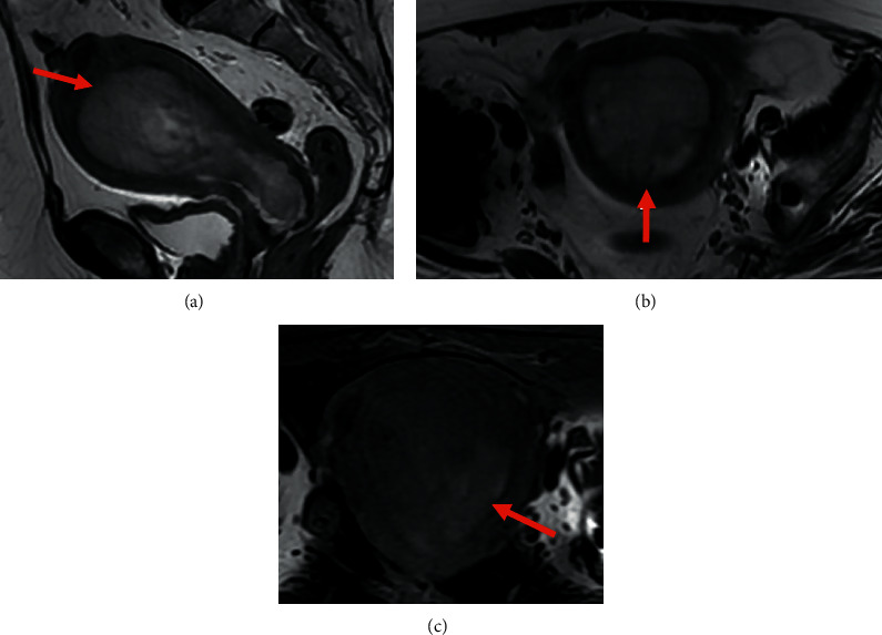
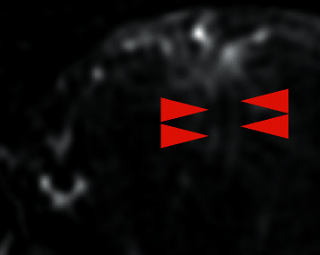
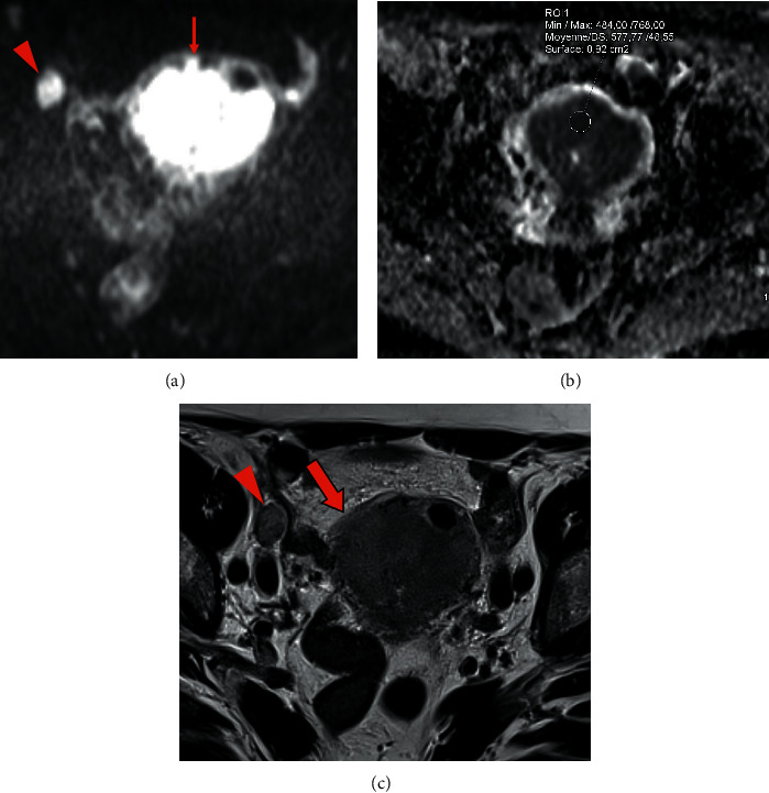
Results: According to their locations, two radiological types of sarcomas were identified: type 1: intracavitary (ESS, MMMT) and type 2: intramyometrial (LMS, STUMP). In both types, all tumors displayed intermediate T2-weighted signal (p < 0.001) and high diffusion-weighted imaging (DWI) b1000 signal (p < 0.001). Dynamic contrast-enhanced (DCE) MRI showed intratumoral pathologic vessels (98%) and heterogeneity at venous phase (p < 0.001). In the type 1 subgroup, all tumors displayed local spread: invasion of junctional zone on T2-weighted imaging (T2WI), irregular margins on DWI, and disruption of arcuate arteries subendometrial ring on DCE-MRI. In the type 2 subgroup, all tumors displayed irregular margins on T2WI, DWI, and DCE-MRI. Tumor heterogeneity was due to necrosis (p < 0.001). Most commonly the tumor was single (61%). In both types, apparent diffusion coefficient (ADC) lesser than or equal to 0.86 × 10⁻³ mm2/s (sensitivity = 73%, specificity = 92%) was suggestive of malignancy.
Conclusion: It may be feasible to get close to histological type of a uterine sarcoma based on our topographic classification into two radiological subgroups, corresponding to two kinds of diagnostic difficulties. Advances in knowledge. MRI signs suggestive of histopathological malignancy are identifiable, considering the triad T2WI/DWI/DCE-MRI, easily for type 1 but less easily for type 2; the threshold value for ADC is 0.86 × 10⁻³ mm2/s.
SarcomaMedicine-Radiology, Nuclear Medicine and Imaging
CiteScore
5.00
自引率
0.00%
发文量
15
审稿时长
14 weeks
期刊介绍:
Sarcoma is dedicated to publishing papers covering all aspects of connective tissue oncology research. It brings together work from scientists and clinicians carrying out a broad range of research in this field, including the basic sciences, molecular biology and pathology and the clinical sciences of epidemiology, surgery, radiotherapy and chemotherapy. High-quality papers concerning the entire range of bone and soft tissue sarcomas in both adults and children, including Kaposi"s sarcoma, are published as well as preclinical and animal studies. This journal provides a central forum for the description of advances in diagnosis, assessment and treatment of this rarely seen, but often mismanaged, group of patients.


 分享
分享
 求助内容:
求助内容: 应助结果提醒方式:
 扫码关注我们
