Juan Hao, Lingjin Liu, Ziqian Liu, Gege Chen, Yunzhao Xiong, Xiangting Wang, Xuelian Ma, Qingyou Xu
{"title":"Aldosterone Induces the Proliferation of Renal Tubular Epithelial Cells <i>In Vivo</i> but Not <i>In Vitro</i>.","authors":"Juan Hao, Lingjin Liu, Ziqian Liu, Gege Chen, Yunzhao Xiong, Xiangting Wang, Xuelian Ma, Qingyou Xu","doi":"10.1155/2021/9943848","DOIUrl":null,"url":null,"abstract":"<p><strong>Objective: </strong>To investigate the proliferation effect of aldosterone on renal tubular epithelial cells <i>in vivo</i> and <i>in vitro</i>.</p><p><strong>Methods: </strong>Thirty-two male C57BL/6J mice (20-22 g) were divided randomly into four groups: sham, unilateral nephrectomy (UN), unilateral nephrectomy plus aldosterone infusion (UA), and UA plus eplerenone (UAE). The kidneys were removed 6 weeks after treatment. Expression of proliferating cell nuclear antigen (PCNA) was detected by immunohistochemistry and western blotting. Human kidney proximal tubular epithelial (HK2) and mouse distal convoluted tubule (mDCT) cell lines were stimulated by aldosterone (0, 10<sup>-9</sup>, 10<sup>-8</sup>, 10<sup>-7</sup>, and 10<sup>-6</sup> mol/L) <i>in vitro</i>. Cells were collected after 3, 6, 12, 24, 36, and 48 h, and proliferation of each group detected by western blotting, flow cytometry, live imaging, and the MTT assay. In addition, mDCT cells were costimulated with a medium containing a final concentration of 161 mmol/L Na<sup>+</sup> and different concentrations of aldosterone, and the number of cells and cellular DNA content was measured by the MTT assay and flow cytometry.</p><p><strong>Results: </strong>Aldosterone could induce a significant increase in the number of PCNA-positive cells in mouse kidneys accompanied by increased deposition of collagen fibers. Eplerenone could inhibit aldosterone-induced cell proliferation and collagen deposition. HK2 cells and mDCT cells administered different concentrations, and different times of aldosterone stimulation failed to cause cell proliferation, and costimulation of aldosterone and salt did not cause proliferation changes in mDCT cells.</p><p><strong>Conclusions: </strong>Aldosterone perfusion can induce proliferation of mouse kidney cells <i>in vivo</i>, and eplerenone can inhibit this change, but aldosterone stimulates HK2 cells and mDCT <i>in vitro</i> without causing their proliferation.</p>","PeriodicalId":2,"journal":{"name":"ACS Applied Bio Materials","volume":" ","pages":"9943848"},"PeriodicalIF":4.7000,"publicationDate":"2021-07-26","publicationTypes":"Journal Article","fieldsOfStudy":null,"isOpenAccess":false,"openAccessPdf":"https://www.ncbi.nlm.nih.gov/pmc/articles/PMC8337160/pdf/","citationCount":"0","resultStr":null,"platform":"Semanticscholar","paperid":null,"PeriodicalName":"ACS Applied Bio Materials","FirstCategoryId":"3","ListUrlMain":"https://doi.org/10.1155/2021/9943848","RegionNum":0,"RegionCategory":null,"ArticlePicture":[],"TitleCN":null,"AbstractTextCN":null,"PMCID":null,"EPubDate":"2021/1/1 0:00:00","PubModel":"eCollection","JCR":"Q2","JCRName":"MATERIALS SCIENCE, BIOMATERIALS","Score":null,"Total":0}
引用次数: 0
Abstract
Objective: To investigate the proliferation effect of aldosterone on renal tubular epithelial cells in vivo and in vitro.
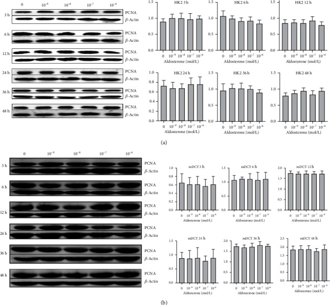
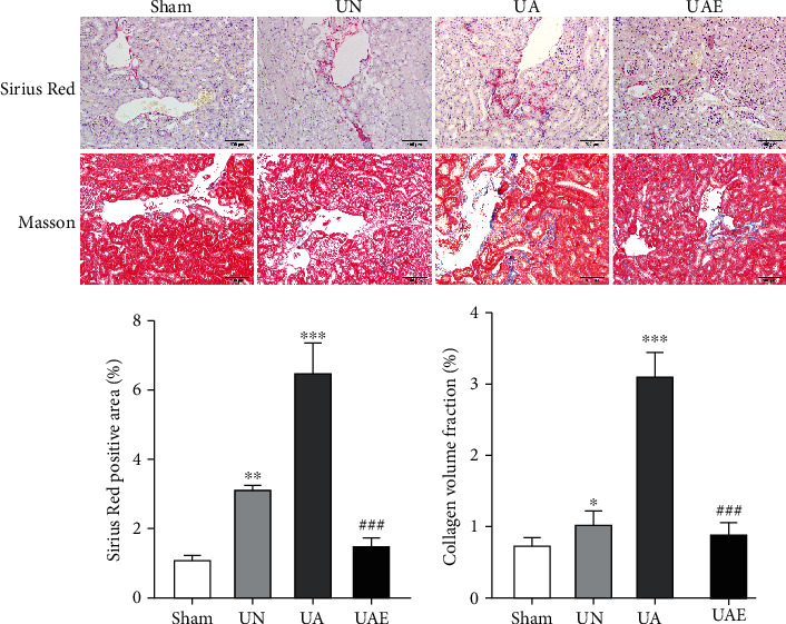
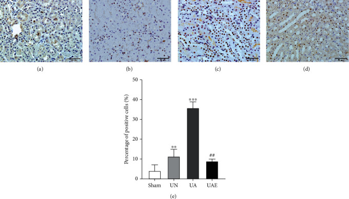
Methods: Thirty-two male C57BL/6J mice (20-22 g) were divided randomly into four groups: sham, unilateral nephrectomy (UN), unilateral nephrectomy plus aldosterone infusion (UA), and UA plus eplerenone (UAE). The kidneys were removed 6 weeks after treatment. Expression of proliferating cell nuclear antigen (PCNA) was detected by immunohistochemistry and western blotting. Human kidney proximal tubular epithelial (HK2) and mouse distal convoluted tubule (mDCT) cell lines were stimulated by aldosterone (0, 10-9, 10-8, 10-7, and 10-6 mol/L) in vitro. Cells were collected after 3, 6, 12, 24, 36, and 48 h, and proliferation of each group detected by western blotting, flow cytometry, live imaging, and the MTT assay. In addition, mDCT cells were costimulated with a medium containing a final concentration of 161 mmol/L Na+ and different concentrations of aldosterone, and the number of cells and cellular DNA content was measured by the MTT assay and flow cytometry.
Results: Aldosterone could induce a significant increase in the number of PCNA-positive cells in mouse kidneys accompanied by increased deposition of collagen fibers. Eplerenone could inhibit aldosterone-induced cell proliferation and collagen deposition. HK2 cells and mDCT cells administered different concentrations, and different times of aldosterone stimulation failed to cause cell proliferation, and costimulation of aldosterone and salt did not cause proliferation changes in mDCT cells.
Conclusions: Aldosterone perfusion can induce proliferation of mouse kidney cells in vivo, and eplerenone can inhibit this change, but aldosterone stimulates HK2 cells and mDCT in vitro without causing their proliferation.
期刊介绍:
ACS Applied Bio Materials is an interdisciplinary journal publishing original research covering all aspects of biomaterials and biointerfaces including and beyond the traditional biosensing, biomedical and therapeutic applications.
The journal is devoted to reports of new and original experimental and theoretical research of an applied nature that integrates knowledge in the areas of materials, engineering, physics, bioscience, and chemistry into important bio applications. The journal is specifically interested in work that addresses the relationship between structure and function and assesses the stability and degradation of materials under relevant environmental and biological conditions.


 分享
分享
 求助内容:
求助内容: 应助结果提醒方式:
 扫码关注我们
