Quantitative dSTORM super-resolution microscopy localizes Aurora kinase A/AURKA in the mitochondrial matrix

IF 4.6 Q2 MATERIALS SCIENCE, BIOMATERIALS ACS Applied Bio Materials Pub Date : 2021-08-31 DOI:10.1111/boc.202100021
Béatrice Durel, Charles Kervrann, Giulia Bertolin
{"title":"Quantitative dSTORM super-resolution microscopy localizes Aurora kinase A/AURKA in the mitochondrial matrix","authors":"Béatrice Durel,&nbsp;Charles Kervrann,&nbsp;Giulia Bertolin","doi":"10.1111/boc.202100021","DOIUrl":null,"url":null,"abstract":"<div>\n \n \n <section>\n \n <h3> Background information</h3>\n \n <p>Mitochondria are dynamic organelles playing essential metabolic and signaling functions in cells. Their ultrastructure has largely been investigated with electron microscopy (EM) techniques. However, quantifying protein-protein proximities using EM is extremely challenging. Super-resolution microscopy techniques as direct stochastic optical reconstruction microscopy (dSTORM) now provide a fluorescent-based, quantitative alternative to EM. Recently, super-resolution microscopy approaches including dSTORM led to valuable advances in our knowledge of mitochondrial ultrastructure, and in linking it with new insights in organelle functions. Nevertheless, dSTORM is mostly used to image integral mitochondrial proteins, and there is little or no information on proteins transiently present at this compartment. The cancer-related Aurora kinase A/AURKA is a protein localized at various subcellular locations, including mitochondria.</p>\n </section>\n \n <section>\n \n <h3> Results</h3>\n \n <p>We first demonstrate that dSTORM coupled to GcoPS can resolve protein proximities within individual submitochondrial compartments. Then, we show that dSTORM provides sufficient spatial resolution to visualize and quantify the most abundant pool of endogenous AURKA in the mitochondrial matrix, as previously shown for overexpressed AURKA. In addition, we uncover a smaller pool of AURKA localized at the OMM, which could have a potential functional readout. We conclude by demonstrating that aldehyde-based fixatives are more specific for the OMM pool of the kinase instead.</p>\n </section>\n \n <section>\n \n <h3> Conclusions</h3>\n \n <p>Our results indicate that dSTORM coupled to GcoPS colocalization analysis is a suitable approach to explore the compartmentalization of non-integral mitochondrial proteins as AURKA, in a qualitative and quantitative manner. This method also opens up the possibility of analyzing the proximity between AURKA and its multiple mitochondrial partners with exquisite spatial resolution, thereby allowing novel insights into the mitochondrial functions controlled by AURKA.</p>\n </section>\n \n <section>\n \n <h3> Significance</h3>\n \n <p>Probing and quantifying the presence of endogenous AURKA – a cell cycle-related protein localized at mitochondria – in the different organelle subcompartments, using quantitative dSTORM super-resolution microscopy.</p>\n </section>\n </div>","PeriodicalId":2,"journal":{"name":"ACS Applied Bio Materials","volume":null,"pages":null},"PeriodicalIF":4.6000,"publicationDate":"2021-08-31","publicationTypes":"Journal Article","fieldsOfStudy":null,"isOpenAccess":false,"openAccessPdf":"https://sci-hub-pdf.com/10.1111/boc.202100021","citationCount":"1","resultStr":null,"platform":"Semanticscholar","paperid":null,"PeriodicalName":"ACS Applied Bio Materials","FirstCategoryId":"99","ListUrlMain":"https://onlinelibrary.wiley.com/doi/10.1111/boc.202100021","RegionNum":0,"RegionCategory":null,"ArticlePicture":[],"TitleCN":null,"AbstractTextCN":null,"PMCID":null,"EPubDate":"","PubModel":"","JCR":"Q2","JCRName":"MATERIALS SCIENCE, BIOMATERIALS","Score":null,"Total":0}
引用次数: 1

Abstract

Background information

Mitochondria are dynamic organelles playing essential metabolic and signaling functions in cells. Their ultrastructure has largely been investigated with electron microscopy (EM) techniques. However, quantifying protein-protein proximities using EM is extremely challenging. Super-resolution microscopy techniques as direct stochastic optical reconstruction microscopy (dSTORM) now provide a fluorescent-based, quantitative alternative to EM. Recently, super-resolution microscopy approaches including dSTORM led to valuable advances in our knowledge of mitochondrial ultrastructure, and in linking it with new insights in organelle functions. Nevertheless, dSTORM is mostly used to image integral mitochondrial proteins, and there is little or no information on proteins transiently present at this compartment. The cancer-related Aurora kinase A/AURKA is a protein localized at various subcellular locations, including mitochondria.

Results

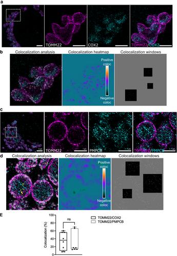
We first demonstrate that dSTORM coupled to GcoPS can resolve protein proximities within individual submitochondrial compartments. Then, we show that dSTORM provides sufficient spatial resolution to visualize and quantify the most abundant pool of endogenous AURKA in the mitochondrial matrix, as previously shown for overexpressed AURKA. In addition, we uncover a smaller pool of AURKA localized at the OMM, which could have a potential functional readout. We conclude by demonstrating that aldehyde-based fixatives are more specific for the OMM pool of the kinase instead.

Conclusions

Our results indicate that dSTORM coupled to GcoPS colocalization analysis is a suitable approach to explore the compartmentalization of non-integral mitochondrial proteins as AURKA, in a qualitative and quantitative manner. This method also opens up the possibility of analyzing the proximity between AURKA and its multiple mitochondrial partners with exquisite spatial resolution, thereby allowing novel insights into the mitochondrial functions controlled by AURKA.

Significance

Probing and quantifying the presence of endogenous AURKA – a cell cycle-related protein localized at mitochondria – in the different organelle subcompartments, using quantitative dSTORM super-resolution microscopy.
Abstract Image

查看原文
分享 分享
微信好友 朋友圈 QQ好友 复制链接
本刊更多论文
定量dSTORM超分辨率显微镜定位线粒体基质中的极光激酶A/AURKA
线粒体是细胞中具有重要代谢和信号功能的动态细胞器。用电子显微镜(EM)技术研究了它们的超微结构。然而,使用EM定量蛋白质-蛋白质接近度是极具挑战性的。超分辨率显微镜技术,如直接随机光学重建显微镜(dSTORM),现在为EM提供了一种基于荧光的定量替代方法。最近,包括dSTORM在内的超分辨率显微镜方法在我们对线粒体超微结构的了解方面取得了有价值的进展,并将其与细胞器功能的新见解联系起来。然而,dSTORM主要用于成像完整的线粒体蛋白质,并且很少或没有关于瞬时存在于该隔室的蛋白质的信息。与癌症相关的极光激酶A/AURKA是一种定位于各种亚细胞位置的蛋白质,包括线粒体。我们首先证明了dSTORM与GcoPS结合可以解决单个亚线粒体区室内的蛋白质接近性。然后,我们证明dSTORM提供了足够的空间分辨率来可视化和量化线粒体基质中最丰富的内源性AURKA库,如先前对过表达的AURKA所示。此外,我们在OMM发现了一个较小的AURKA池,它可能具有潜在的功能读数。我们通过证明醛基固定剂对激酶的OMM池更具特异性而得出结论。我们的研究结果表明,dSTORM结合GcoPS共定位分析是一种合适的方法,可以定性和定量地探索非完整线粒体蛋白作为AURKA的区区化。这种方法也为分析AURKA及其多个线粒体伴侣之间的接近性提供了可能性,具有精细的空间分辨率,从而对AURKA控制的线粒体功能有了新的认识。意义:利用dSTORM超分辨率显微镜检测和量化不同细胞器亚室中内源性AURKA(一种定位于线粒体的细胞周期相关蛋白)的存在。
本文章由计算机程序翻译,如有差异,请以英文原文为准。
求助全文
约1分钟内获得全文 去求助
来源期刊
ACS Applied Bio Materials
ACS Applied Bio Materials Chemistry-Chemistry (all)
CiteScore
9.40
自引率
2.10%
发文量
464
期刊最新文献
A Systematic Review of Sleep Disturbance in Idiopathic Intracranial Hypertension. Advancing Patient Education in Idiopathic Intracranial Hypertension: The Promise of Large Language Models. Anti-Myelin-Associated Glycoprotein Neuropathy: Recent Developments. Approach to Managing the Initial Presentation of Multiple Sclerosis: A Worldwide Practice Survey. Association Between LACE+ Index Risk Category and 90-Day Mortality After Stroke.
×
引用
GB/T 7714-2015
复制
MLA
复制
APA
复制
导出至
BibTeX EndNote RefMan NoteFirst NoteExpress
×
×
提示
您的信息不完整,为了账户安全,请先补充。
现在去补充
×
提示
您因"违规操作"
具体请查看互助需知
我知道了
×
提示
现在去查看 取消
×
提示
确定
0
微信
客服QQ
Book学术公众号 扫码关注我们
反馈
×
意见反馈
请填写您的意见或建议
请填写您的手机或邮箱
已复制链接
已复制链接
快去分享给好友吧!
我知道了
×
扫码分享
扫码分享
Book学术官方微信
Book学术文献互助
Book学术文献互助群
群 号:481959085
Book学术
文献互助 智能选刊 最新文献 互助须知 联系我们:info@booksci.cn
Book学术提供免费学术资源搜索服务,方便国内外学者检索中英文文献。致力于提供最便捷和优质的服务体验。
Copyright © 2023 Book学术 All rights reserved.
ghs 京公网安备 11010802042870号 京ICP备2023020795号-1