Comparative proteome analysis of form-deprivation myopia in sclera with iTRAQ-based quantitative proteomics.
IF 1.4 3区 医学Q4 BIOCHEMISTRY & MOLECULAR BIOLOGYMolecular VisionPub Date : 2021-09-01eCollection Date: 2021-01-01
Ying Yuan, Chengcheng Zhu, Mingming Liu, Bilian Ke
{"title":"Comparative proteome analysis of form-deprivation myopia in sclera with iTRAQ-based quantitative proteomics.","authors":"Ying Yuan, Chengcheng Zhu, Mingming Liu, Bilian Ke","doi":"","DOIUrl":null,"url":null,"abstract":"<p><strong>Objective: </strong>Scleral remodeling plays a key role in axial elongation in myopia. The aim of the present study was to identify the proteomics changes and specific signaling networks to gain insight into the molecular basis of scleral remodeling in myopic eyes.</p><p><strong>Methods: </strong>Guinea pig form-deprivation myopia was induced with a translucent diffuser on a random eye for 4 weeks, while the other eye served as the contralateral control group. The axial length and refraction were measured at the beginning and end of the treatment. The proteins were extracted from the sclerae of each group and prepared for quantitative isobaric tags for relative and absolute quantification (iTRAQ) labeling combined with liquid chromatography-tandem mass spectrometry (LC-MS/MS) analysis. The coexpression networks and protein functions were analyzed using Gene Ontology (GO) and Ingenuity Pathway Analysis (IPA). Quantitative real-time PCR (qRT-PCR) and western blotting were performed to confirm the authenticity and accuracy of the iTRAQ results.</p><p><strong>Results: </strong>After 4 weeks, the form-deprivation eyes developed significant degrees of myopia, and the axial length increased statistically significantly (p<0.05). A total of 2,579 unique proteins with <1% false discovery rate (FDR) were identified. Furthermore, 56 proteins were found to be upregulated, and 84 proteins were found to be downregulated, with a threshold of a 1.2-fold change and p<0.05 in the myopia group, when compared to the control group. Further bioinformatics analysis indicated that 44 of 140 differentially expressed proteins were involved in cellular movement and cellular assembly and organization. The qRT-PCR or western blotting results confirmed that myosin IIB, ACTIN3, and cellular cytoskeletons were downregulated, while RhoA and RAP1A were upregulated in the sclera in myopic eyes. These results were consistent with the proteomics results.</p><p><strong>Conclusions: </strong>Proteomics and bioinformatics results can be helpful for identifying proteins and providing new insights for better understanding of the molecular mechanism underlying scleral remodeling. These results revealed that the proteins associated with cellular movement and cellular assembly and organization are altered during the development of myopia. Furthermore, RhoA plays a key role in the pathways involved in cellular movement and cellular assembly and organization.</p>","PeriodicalId":18866,"journal":{"name":"Molecular Vision","volume":"27 ","pages":"494-505"},"PeriodicalIF":1.4000,"publicationDate":"2021-09-01","publicationTypes":"Journal Article","fieldsOfStudy":null,"isOpenAccess":false,"openAccessPdf":"https://ftp.ncbi.nlm.nih.gov/pub/pmc/oa_pdf/6f/ad/mv-v27-494.PMC8410231.pdf","citationCount":"0","resultStr":null,"platform":"Semanticscholar","paperid":null,"PeriodicalName":"Molecular Vision","FirstCategoryId":"3","ListUrlMain":"","RegionNum":3,"RegionCategory":"医学","ArticlePicture":[],"TitleCN":null,"AbstractTextCN":null,"PMCID":null,"EPubDate":"2021/1/1 0:00:00","PubModel":"eCollection","JCR":"Q4","JCRName":"BIOCHEMISTRY & MOLECULAR BIOLOGY","Score":null,"Total":0}
引用次数: 0
Abstract
Objective: Scleral remodeling plays a key role in axial elongation in myopia. The aim of the present study was to identify the proteomics changes and specific signaling networks to gain insight into the molecular basis of scleral remodeling in myopic eyes.
Methods: Guinea pig form-deprivation myopia was induced with a translucent diffuser on a random eye for 4 weeks, while the other eye served as the contralateral control group. The axial length and refraction were measured at the beginning and end of the treatment. The proteins were extracted from the sclerae of each group and prepared for quantitative isobaric tags for relative and absolute quantification (iTRAQ) labeling combined with liquid chromatography-tandem mass spectrometry (LC-MS/MS) analysis. The coexpression networks and protein functions were analyzed using Gene Ontology (GO) and Ingenuity Pathway Analysis (IPA). Quantitative real-time PCR (qRT-PCR) and western blotting were performed to confirm the authenticity and accuracy of the iTRAQ results.
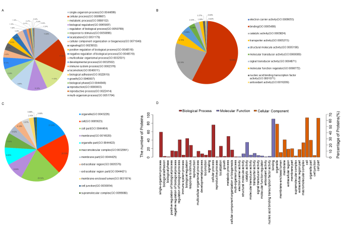
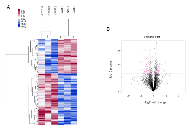
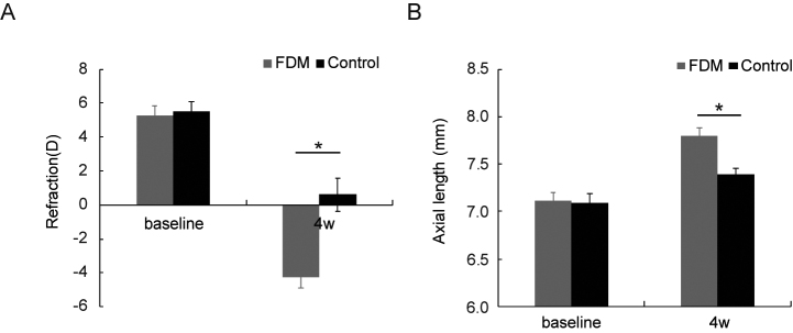
Results: After 4 weeks, the form-deprivation eyes developed significant degrees of myopia, and the axial length increased statistically significantly (p<0.05). A total of 2,579 unique proteins with <1% false discovery rate (FDR) were identified. Furthermore, 56 proteins were found to be upregulated, and 84 proteins were found to be downregulated, with a threshold of a 1.2-fold change and p<0.05 in the myopia group, when compared to the control group. Further bioinformatics analysis indicated that 44 of 140 differentially expressed proteins were involved in cellular movement and cellular assembly and organization. The qRT-PCR or western blotting results confirmed that myosin IIB, ACTIN3, and cellular cytoskeletons were downregulated, while RhoA and RAP1A were upregulated in the sclera in myopic eyes. These results were consistent with the proteomics results.
Conclusions: Proteomics and bioinformatics results can be helpful for identifying proteins and providing new insights for better understanding of the molecular mechanism underlying scleral remodeling. These results revealed that the proteins associated with cellular movement and cellular assembly and organization are altered during the development of myopia. Furthermore, RhoA plays a key role in the pathways involved in cellular movement and cellular assembly and organization.
期刊介绍:
Molecular Vision is a peer-reviewed journal dedicated to the dissemination of research results in molecular biology, cell biology, and the genetics of the visual system (ocular and cortical).
Molecular Vision publishes articles presenting original research that has not previously been published and comprehensive articles reviewing the current status of a particular field or topic. Submissions to Molecular Vision are subjected to rigorous peer review. Molecular Vision does NOT publish preprints.
For authors, Molecular Vision provides a rapid means of communicating important results. Access to Molecular Vision is free and unrestricted, allowing the widest possible audience for your article. Digital publishing allows you to use color images freely (and without fees). Additionally, you may publish animations, sounds, or other supplementary information that clarifies or supports your article. Each of the authors of an article may also list an electronic mail address (which will be updated upon request) to give interested readers easy access to authors.
 分享
分享


 求助内容:
求助内容: 应助结果提醒方式:
 扫码关注我们
