Pan He, Hang Qu, Ming Cai, Weijie Liu, Xinyi Gu, Qiang Ma
{"title":"Structural Alteration of Medial Temporal Lobe Subfield in the Amnestic Mild Cognitive Impairment Stage of Alzheimer's Disease.","authors":"Pan He, Hang Qu, Ming Cai, Weijie Liu, Xinyi Gu, Qiang Ma","doi":"10.1155/2022/8461235","DOIUrl":null,"url":null,"abstract":"<p><strong>Objective: </strong>Volume reduction and structural abnormality is the most replicated finding in neuroimaging studies of Alzheimer's disease (AD). Amnestic mild cognitive impairment (aMCI) is the early stage of AD development. Thus, it is necessary to investigate the link between atrophy of regions of interest (ROIs) in medial temporal lobe, the variation trend of ROI densities and volumes among patients with cognitive impairment, and the distribution characteristics of ROIs in the aMCI group, Alzheimer's disease (AD) group, and normal control (NC) group.</p><p><strong>Methods: </strong>30 patients with aMCI, 16 patients with AD, and 30 NC are recruited; magnetic resonance imaging (MRI) brain scans are conducted. Voxel-based morphometry was employed to conduct the quantitative measurement of gray matter densities of the hippocampus, amygdala, entorhinal cortex, and mammillary body (MB). FreeSurfer was utilized to automatically segment the hippocampus into 21 subregions and the amygdala into 9 subregions. Then, their subregion volumes and total volume were calculated. Finally, the ANOVA and multiple comparisons were performed on the above-mentioned data from these three groups.</p><p><strong>Results: </strong>AD had lower GM densities than MCI, and MCI had lower GM densities than NC, but not all of the differences were statistically significant. In the comparisons of AD-aMCI-NC, AD-aMCI, and AD-NC, the hippocampus, amygdala, and entorhinal cortex showed differences in the gray matter densities (<i>p</i> < 0.05); the differences of mammillary body densities were not significant in the random comparison between these three groups (<i>p</i> > 0.05). The hippocampus densities and volumes of the subjects from the aMCI group and the AD group were bilaterally symmetric. The gray matter densities of the right side of the entorhinal cortex inside each group and the hippocampus from the NC group were higher than those of the left side (<i>p</i> < 0.05), and the gray matter densities of the amygdala and mammillary body were bilaterally symmetric in the three groups (<i>p</i> > 0.05). There were no gender differences of four ROIs in the AD, aMCI, and NC groups (<i>p</i> > 0.05). The volume differences of the hippocampus presubiculum-body and parasubiculum manifest no statistical significance (<i>p</i> > 0.05) in the random comparison between these three groups. Volume differences of the left amygdala basal nucleus, the left lateral nucleus, the left cortical amygdala transitional area, the left paravamnion nucleus, and bilateral hippocampal amygdala transition area (HATA) had statistical differences only between the AD group and the NC group (<i>p</i> < 0.05).</p><p><strong>Conclusion: </strong>Structural defects of medial temporal lobe subfields were revealed in the aMCI and AD groups. Decreased gray matter densities of the hippocampus, entorhinal cortex, and amygdala could distinguish patients with early stage of AD between aMCI and NC. Volume decline of the hippocampus and amygdala subfields could only distinguish AD between NC.</p>","PeriodicalId":51299,"journal":{"name":"Neural Plasticity","volume":" ","pages":"8461235"},"PeriodicalIF":3.7000,"publicationDate":"2022-01-24","publicationTypes":"Journal Article","fieldsOfStudy":null,"isOpenAccess":false,"openAccessPdf":"https://www.ncbi.nlm.nih.gov/pmc/articles/PMC8803445/pdf/","citationCount":"5","resultStr":null,"platform":"Semanticscholar","paperid":null,"PeriodicalName":"Neural Plasticity","FirstCategoryId":"3","ListUrlMain":"https://doi.org/10.1155/2022/8461235","RegionNum":4,"RegionCategory":"医学","ArticlePicture":[],"TitleCN":null,"AbstractTextCN":null,"PMCID":null,"EPubDate":"2022/1/1 0:00:00","PubModel":"eCollection","JCR":"Q2","JCRName":"NEUROSCIENCES","Score":null,"Total":0}
引用次数: 5
Abstract
Objective: Volume reduction and structural abnormality is the most replicated finding in neuroimaging studies of Alzheimer's disease (AD). Amnestic mild cognitive impairment (aMCI) is the early stage of AD development. Thus, it is necessary to investigate the link between atrophy of regions of interest (ROIs) in medial temporal lobe, the variation trend of ROI densities and volumes among patients with cognitive impairment, and the distribution characteristics of ROIs in the aMCI group, Alzheimer's disease (AD) group, and normal control (NC) group.
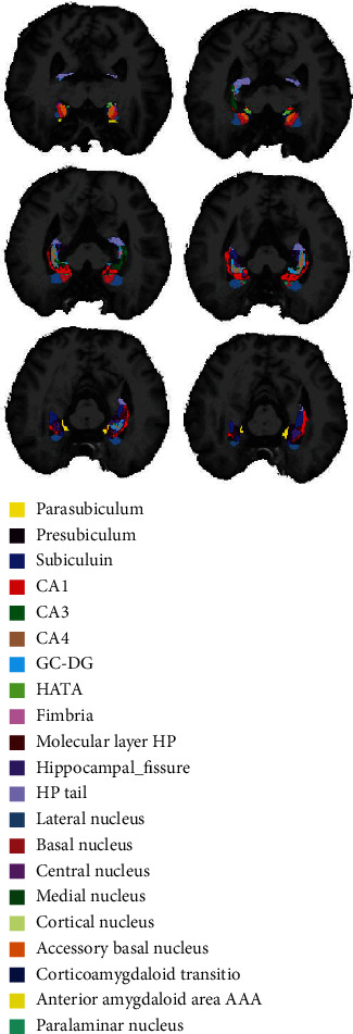
Methods: 30 patients with aMCI, 16 patients with AD, and 30 NC are recruited; magnetic resonance imaging (MRI) brain scans are conducted. Voxel-based morphometry was employed to conduct the quantitative measurement of gray matter densities of the hippocampus, amygdala, entorhinal cortex, and mammillary body (MB). FreeSurfer was utilized to automatically segment the hippocampus into 21 subregions and the amygdala into 9 subregions. Then, their subregion volumes and total volume were calculated. Finally, the ANOVA and multiple comparisons were performed on the above-mentioned data from these three groups.
Results: AD had lower GM densities than MCI, and MCI had lower GM densities than NC, but not all of the differences were statistically significant. In the comparisons of AD-aMCI-NC, AD-aMCI, and AD-NC, the hippocampus, amygdala, and entorhinal cortex showed differences in the gray matter densities (p < 0.05); the differences of mammillary body densities were not significant in the random comparison between these three groups (p > 0.05). The hippocampus densities and volumes of the subjects from the aMCI group and the AD group were bilaterally symmetric. The gray matter densities of the right side of the entorhinal cortex inside each group and the hippocampus from the NC group were higher than those of the left side (p < 0.05), and the gray matter densities of the amygdala and mammillary body were bilaterally symmetric in the three groups (p > 0.05). There were no gender differences of four ROIs in the AD, aMCI, and NC groups (p > 0.05). The volume differences of the hippocampus presubiculum-body and parasubiculum manifest no statistical significance (p > 0.05) in the random comparison between these three groups. Volume differences of the left amygdala basal nucleus, the left lateral nucleus, the left cortical amygdala transitional area, the left paravamnion nucleus, and bilateral hippocampal amygdala transition area (HATA) had statistical differences only between the AD group and the NC group (p < 0.05).
Conclusion: Structural defects of medial temporal lobe subfields were revealed in the aMCI and AD groups. Decreased gray matter densities of the hippocampus, entorhinal cortex, and amygdala could distinguish patients with early stage of AD between aMCI and NC. Volume decline of the hippocampus and amygdala subfields could only distinguish AD between NC.
期刊介绍:
Neural Plasticity is an international, interdisciplinary journal dedicated to the publication of articles related to all aspects of neural plasticity, with special emphasis on its functional significance as reflected in behavior and in psychopathology. Neural Plasticity publishes research and review articles from the entire range of relevant disciplines, including basic neuroscience, behavioral neuroscience, cognitive neuroscience, biological psychology, and biological psychiatry.


 分享
分享
 求助内容:
求助内容: 应助结果提醒方式:
 扫码关注我们
